|
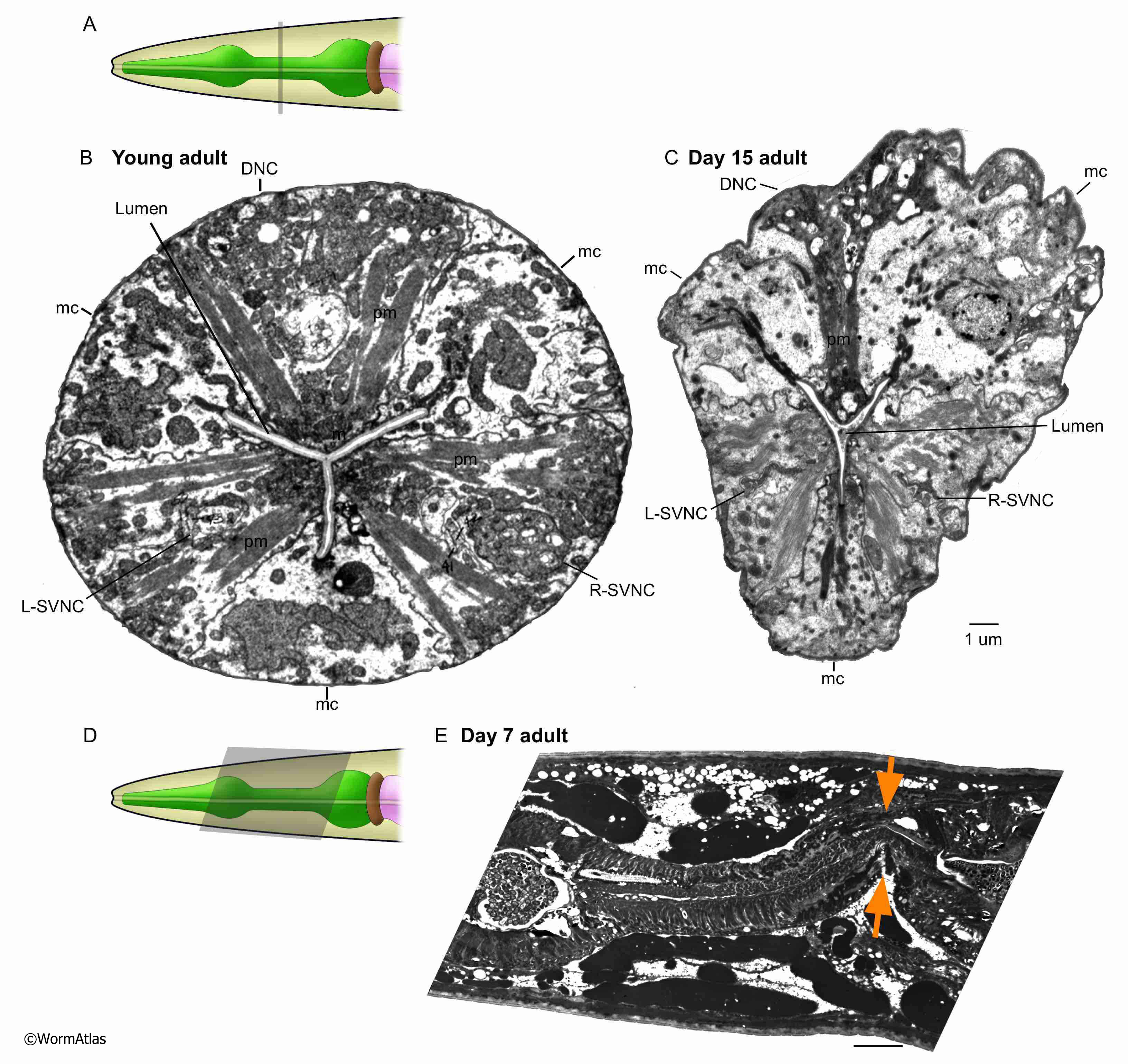
APhaFIG 8: Isthmus deterioration during aging.
A. Diagram showing the approximate positions of B&C in the pharyngeal isthmus.
B. Cross-section from the isthmus region of a young adult pharynx. The pharyngeal muscles (pm) and marginal cells (C) are triradially arranged around the central lumen, which is compressed in this region. The nerve cords (DNC, L&R-SVNC) within the pharynx muscles are different in each segment in this adult. (Image source: N2T [N. Thomson] A104-11.)
C. Cross-section of the isthmus region in this 15-day-old adult showing deterioration of the dorsal pharyngeal muscle and all three of the nerve cords. Note that only the dorsal muscle shows flagrant shape change here, as well as increased cytoplasmic staining, while the two subcventral muscles retain more normal structures. The marginal cells appear intact but disorganized. The lumen remains compressed but contains debris. Bar, 1 micron. (Image source: N813 [D. Hall] G531.)
D. Diagram of C. elegans pharynx with grey shape showing the approximate position of E.
E. Lengthwise section of the pharyngeal isthmus from this 7-day adult shows weakening and kinking of the structure (orange arrows). (Image source: N824 [D. Hall] N4924.) Bar, 5 microns.
Click on picture for full resolution image.
|