|
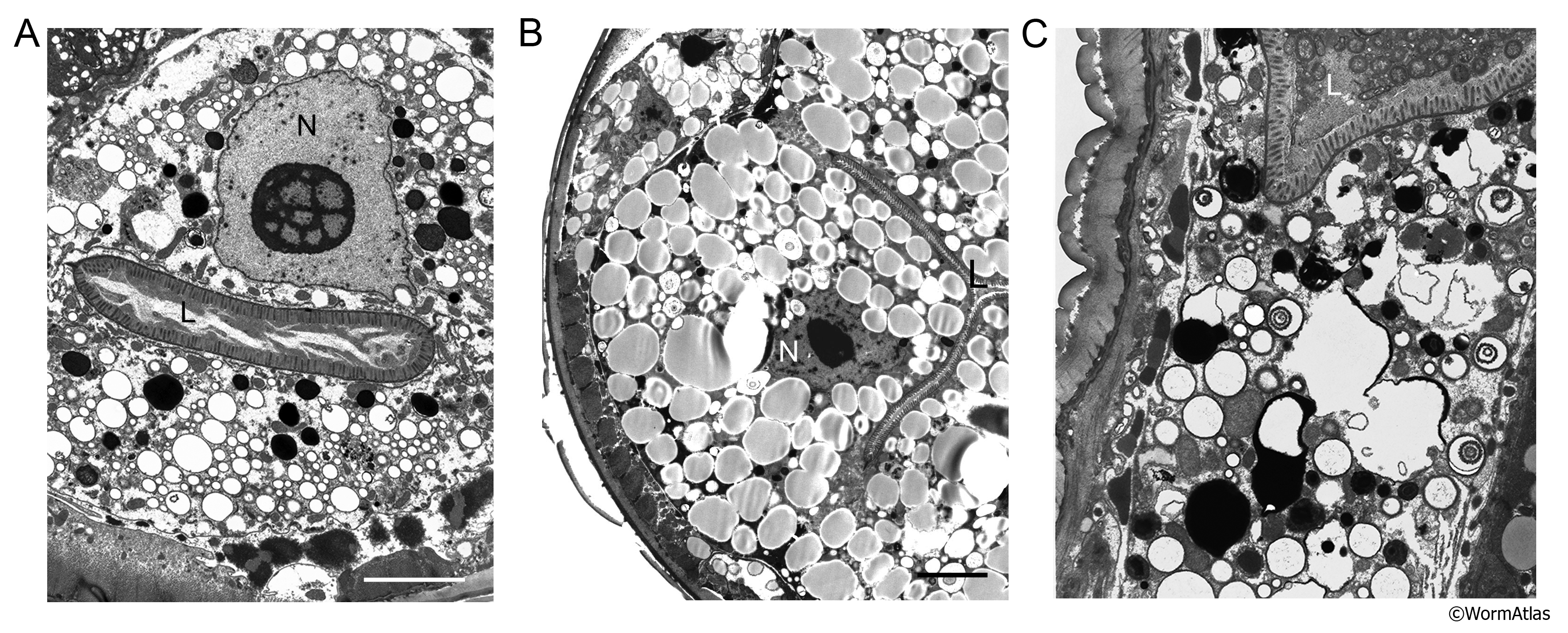
AIntFIG 7: Changes in intestinal cell cytoplasm.
A. In a day 7 adult intestinal cell, the nucleus (N) has lost heterochromatin and contains an enlarged, vacuolated nucleolus. The cytoplasm is highly vacuolated, with a profound reduction in ground substance and RER, although many mitochondria remain intact. The apical zones are studded with intact microvilli but the lumen (L) is almost empty. (Image source: N826 [Hall] 5353.) Bar, 5 µm.
B. A day 15 adult (class A) intestinal cell in which the cytoplasm is choked by lipid storage droplets, having moderately electron dense interiors. The nucleus appears small and more electron dense, seeming to have shrunken in diameter, though this thin section may not pass through the center of the nucleus. L, lumen; N, nucleus. (Image source: N812 [Hall] F815.) Bar, 5 µm.
C. A day 15 adult (class B) intestinal cell showing significant breakdown of the cytoplasmic components. Whorl like inclusions can be seen in some places, and most vacuoles are larger in diameter and more empty of contents compared to the other examples here. (Image source: N815 [Hall] G989.)
Click on picture for full resolution image.
|