|
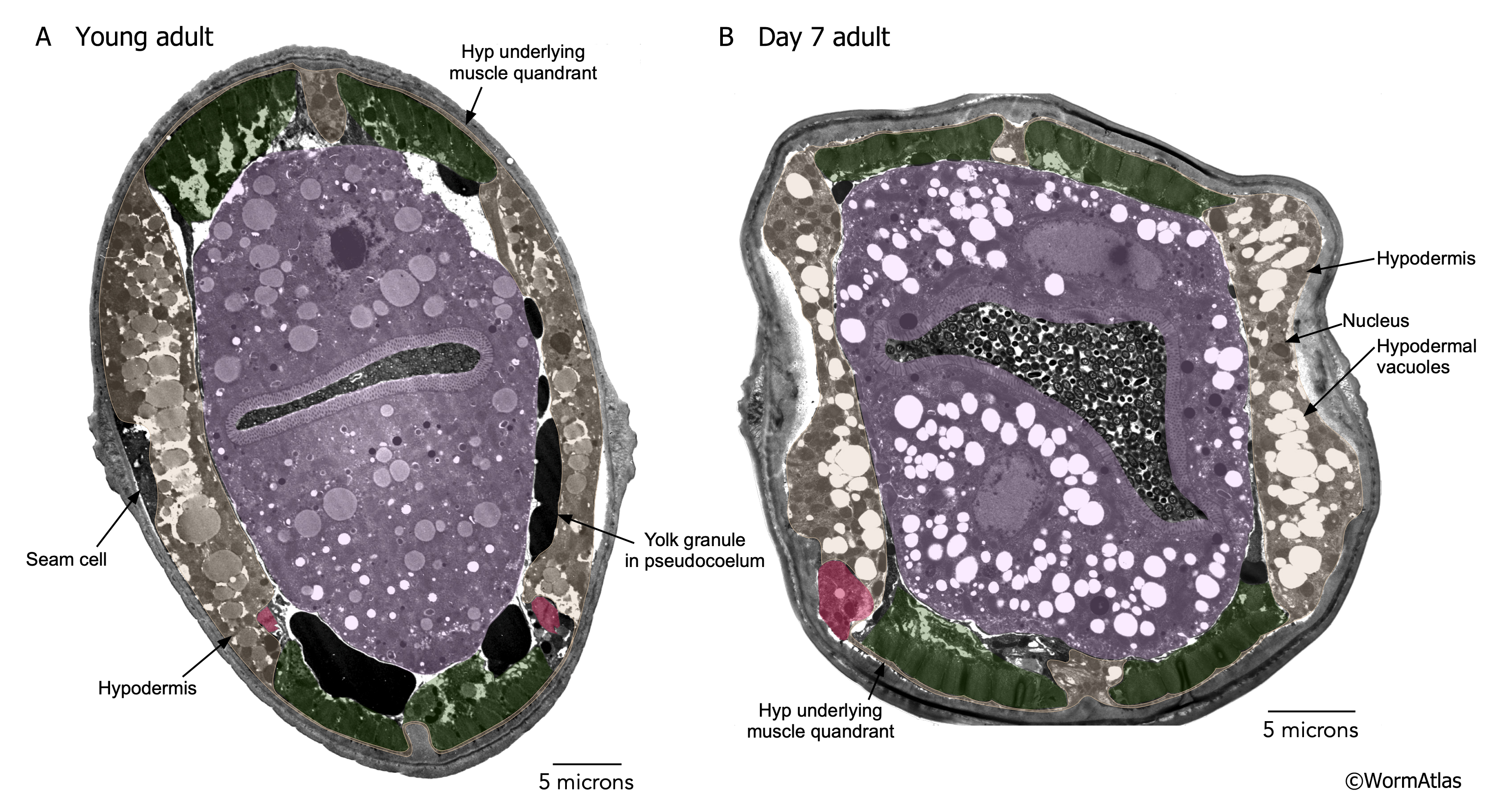
AHypFIG 3: Vacuoles appear in aging hypodermis.
A. Cross-section of young adult from the midbody region, outside of the region of the gonad arms. The hyp (tan) is most evident on the lateral sides of the body adjacent to the seam cells. As in previous panels above, the seam cell is only sometimes evident within the lateral hypodermis, as marked on the right side of the adult. In this individual, the hypodermal cytoplasm appears to contain large droplets of fatty material. Excretory cell (magenta). Bar, 5 microns.
B. Cross section of a day 7 adult. At this age, large numbers of empty vacuoles have appeared in the hypodermis, replacing the droplets in the young adult with similar vacuoles also appearing in the intestinal cytoplasm (pink). Muscles (green) still seem well anchored to the cuticle at day 7. Note that the seam cell cytoplasm on the left side is very electron dense, in contrast to the very light staining of the nearby hypodermal cytoplasm. Bar, 5 µm. (Image source: A. N903C X751 [D. Hall]; B. N826 G5719 [D. Hall].)
Click on picture for full resolution image.
|