|
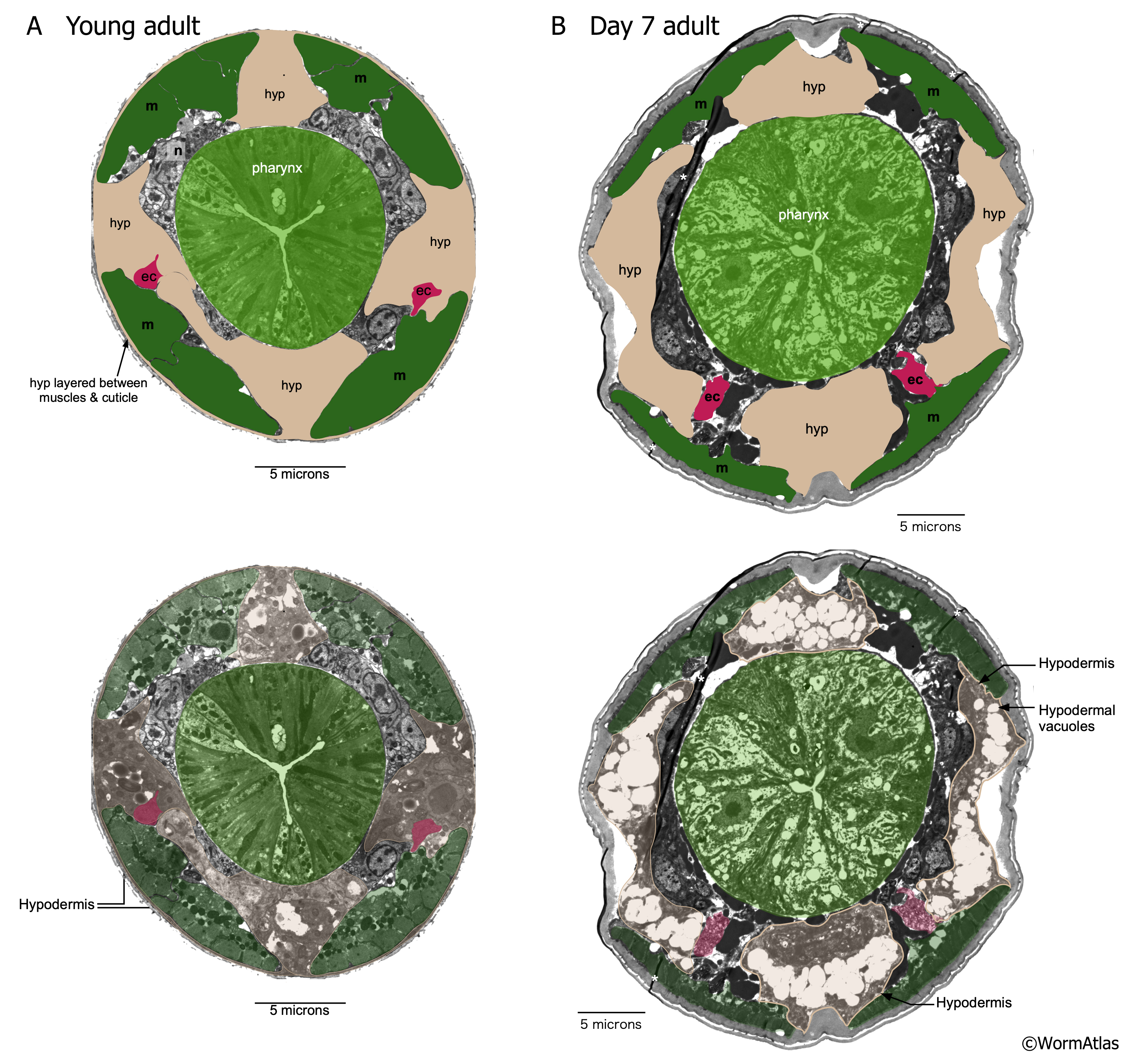
AHypFIG 2: Vacuoles in the head hypodermal cells of older adult.
A. Cross-section of the head region in a young adult with the hypodermal cells indicated with tan shading. In the head, hypodermal cells are not all syncytial. Excretory cell ec (magenta); muscle m (green); Bar, 5 µm.
B. In this day 7 adult, the hypodermal cells (tan) in the head region have accumulated large numbers of vacuoles but retain the same approximate body volume as in the young adult. Overall diameter of the body is now larger, and the body is also longer, so total body volume is increased. Neither seam cell is now evident, possibly due to similar changes inside that tissue, or due to the distortions of the lateral hypodermis by vacuolation. There also appears to be some separation of the hypodermis from the cuticle attachments along several regions of the body, but muscles (green) still seem well anchored to the cuticle. Asterisks indicate image artefacts not indicative of body morphology. Bar, 5 µm. (Image sources: A. N510C V782 [D. Hall]; B. N825 G4168 [D. Hall].)
Click on picture for full resolution image.
|