|
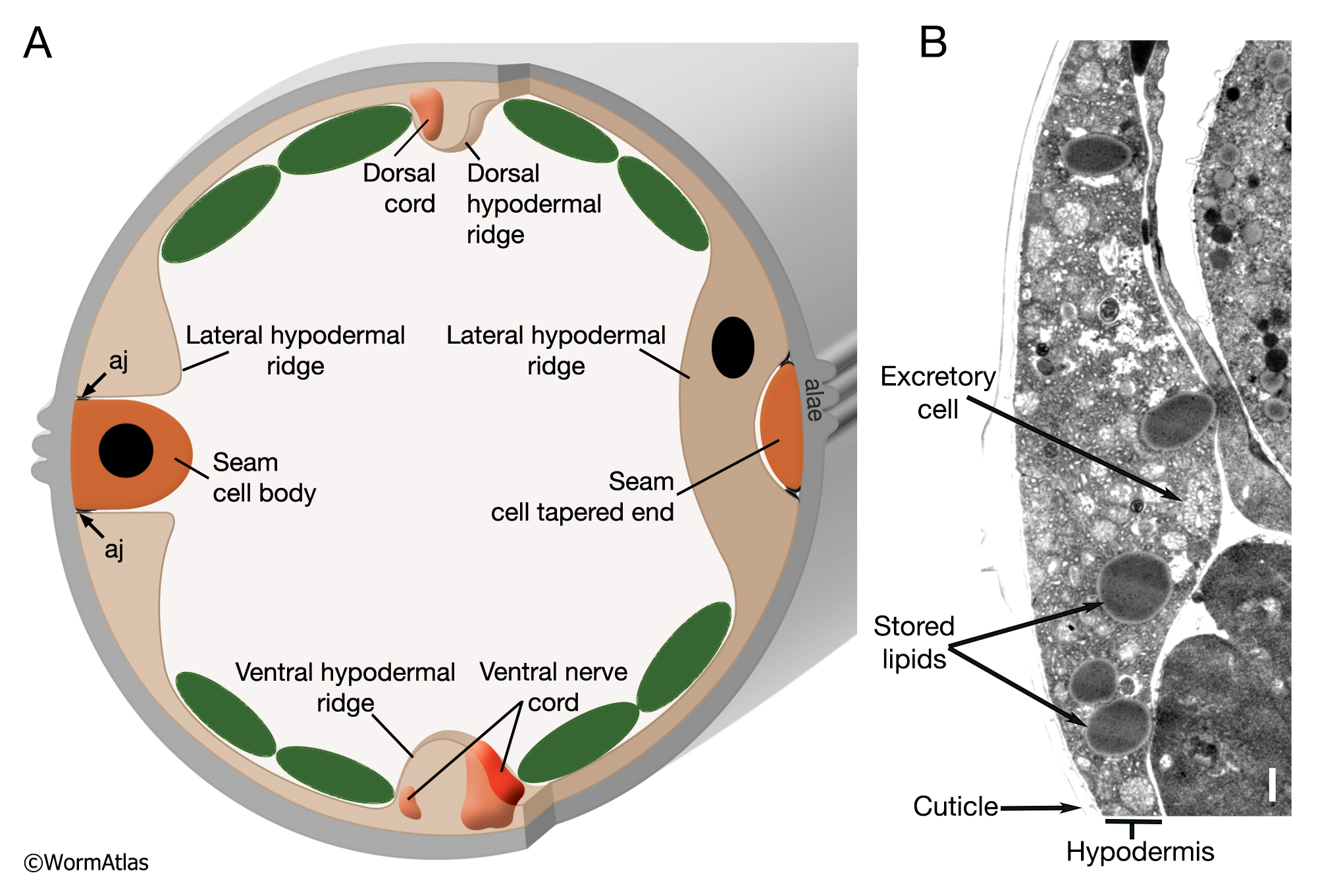
AHypFIG 1: Structure of the hypodermis and surrounding tissues.
A. Schematic showing the interactive structure of the hypodermis with the body wall muscles, seam cells and the cuticle. The hypodermis is composed of four major longitudinal ridges (ventral, dorsal, and left/right lateral) that are joined circumferentially by thin sheets of cytoplasm. At each original cell body of the seam syncytium, where the nuclei are positioned, the seam completely interrupts hyp 7, whereas at the ends of the cells, the seam is so thin that hyp 7 runs behind it, enfolding it completely. At its apical surface, seam makes adherens junctions (aj) to the hypodermis throughout its length. (Green ovals) Muscle quadrants.
B. TEM, transverse section representing an area through the hypodermis in an adult animal. Seam cell is tapered down here and not easily seen. Bar, 1µm. (Image source: [Hall] N506 M700.) (See also Hermaphrodite Hypodermis and Hermaphrodite Seam Cells for more details.)
Click on picture for full resolution image.
|