 Click pictures for higher resolution images Click pictures for higher resolution images
GLR is a set of six cells that are situated posteriorly to the
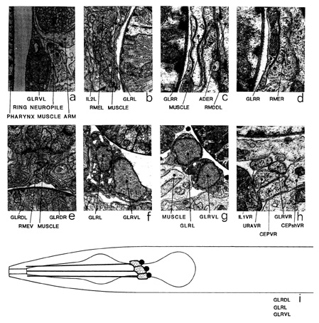
nerve ring, close to the neuropile (a). Cell bodies are situated in a
sixfold symmetrical arrangement and send processes that project
anteriorly and run adjacent to the pharynx. All six processes peter
out and end, with no terminal specializations, at the level of the
junction of the pharynx and the buccal cavity. Each process flattens
out into a sheet, which touches its neighbour on each side, forming a
cylinder as they pass the inside surface of the nerve ring. Muscle
arms from the head muscles run posteriorly down the outside of the
nerve ring and then turn to run anteriorly, next to the inside surface
of the nerve ring, where they are sandwiched between the cylinder of GLR cells and the motor endplate region of the nerve ring (a,
b, c). It is in this region that the muscle arms receive their
synaptic input from the motoneurons of the nerve ring. The muscle
arms are highly ordered in this region, arms from individual muscle
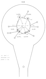
rows running along specific GLR cells (figure 15). Gap junctions are
seen between GLR cells and muscle arms (c) and also RME motoneurons (d), but not between adjacent GLR cells even though
they touch in this region (e). At the anterior extremity of the ring
the GLR processes lose their sheet-like morphology and revert
to a small cylindrical form. These processes run anteriorly and are
always closely apposed with the IL1 (h) dendrites until they
end. The nuclei of the GLR cells are small and the cytoplasm
contains large, irregularly shaped, membrane-bound vesicles, which are
more prominent in the larva (f) than the adult stages (g). No
chemical synapses have been seen either to or from these cells. The
disposition and the layout of these cells suggest that they might be
glial cells and might act to guide growing muscle arms and the sensory
dendrites from each of the labia in the head. Magnifications: (a, f,
g) x 8500, (b, e) x 12750, (c, d, h) x 25500.

|
|

Click pictures for higher resolution images


