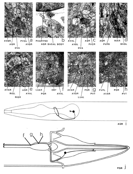
Although AQR and PQR have been given different class names, they have several
features in common and so have been grouped together. Each is derived
from an equivalent position on bilaterally symmetrical lineages
(Sulston & Horvitz 1977) and each has a small cilium, which is not
part or a sensillum but is free in the body cavity (b). The cell body
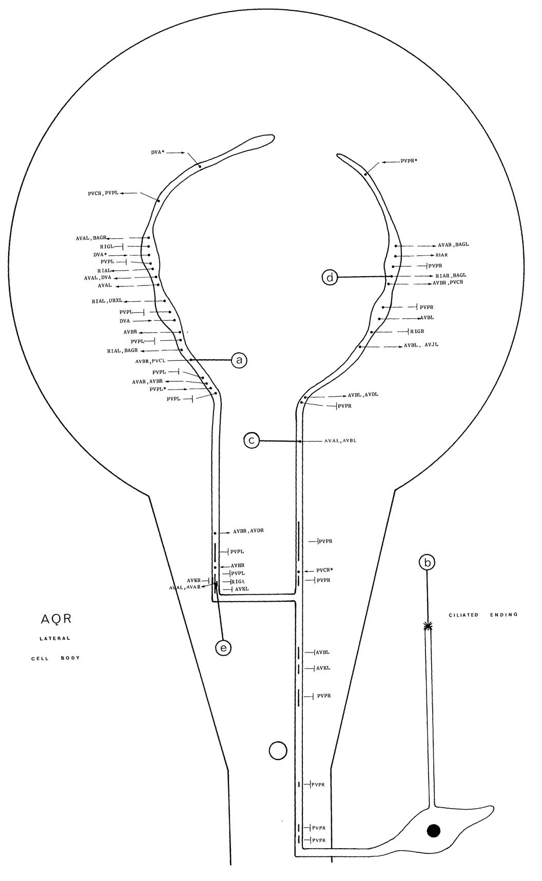
of AQR is situated laterally on the right-hand side near the
posterior bulb of the pharynx. The cilium is on a small process
emanating from the cell body (i). The cell body of PQR is in
the left lumbar ganglion and its cilium is near the end of a
posteriorly directed process (j). The main process of AQR enters the ventral cord via the right-hand deirid commissure and runs
anteriorly. It splits near the nerve ring and the two branches run
round each side or the nerve ring, near the middle orthering neuropile
and in close association with the process of DVA. The
processes or AQR end without meeting near the dorsal mid-line.
The main synaptic output is to AVB (a, c), AVA (c, e), RIA (d), BAG (d), PVC (a) and AVD. AQR has noticeably denser clusters or vesicles presynaptically
than most of the other classes of neuron. There is some synaptic input
from DVA (*c) and many gap junctions to PVP and also
some to AVK (*f) and RIG. PQR sends an
anteriorly directed process that enters the pre-anal ganglion and runs
anteriorly in the ventral region of the process bundle, eventually
ending somewhere in the posterior half or the ventral cord. The main
synaptic output of PQR is directed to AVA (r, g) and AVD (g), usually in dyadic combinations. There are also gap
junctions to PVP (h) and there is some synaptic input from PVN (*c). Magnifications: (a-c, e, f) x 25500, (d) x 12750,
(g, h) x 38250.


Click pictures for higher resolution images