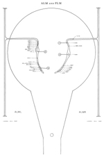
ALM and PLM are two sets of two sensory neurons that transduce touch
stimuli (Chalfie & Sulston 1981). Both ALM and PLM have long lateral
processes, closely apposed to the cuticle, which contain large, darkly
staining microtubules (g) (Chalfie & Thomson 1982). Microtubules
with the same appearance are seen in AVM and PVM, which are also part
of the touch-transducing system. The cell bodies of ALM are
situated laterally in the mid-body (i). Anteriorly directed processes
leave the cell bodies and run near the dorsal edge of the lateral
hypodermal ridges in close association with the processes of ALN (*d).
Each process sends off a branch, which enters the nerve ring
sub-dorsally; this then runs ventrally round the ring near the inside
surface, ending soon after it meets a process of AVM. The processes of ALM are predominantly presynaptic in the nerve ring and synapse onto BDU (a), PVC (b) and CEP (c). There are gap junctions to AVM (*d) and PVR (d). The cell bodies of PLM are situated in the lumbar
ganglia (j). Anteriorly and posteriorly directed processes emanate
from the cell bodies and run near the ventral edge of the lateral
hypodermal ridges (g) in close association with the processes of PLN for part of their length. Gap junctions are made to PVC (h), LUA (*d), and PVR where the processes of PLM cross the lumbar commissures
(j). Each HSN sends out a short ventral branch, which receives a
single synapse from the lateral PLM processes (g). The processes of PLM turn and enter the ventral cord via a commissure near the vulva.
The process of PLML does not get over the hypodermal ridge (which is
rather wide at this point due to the proximity or the vulva) and has
no synapses. The process of PLMR runs along the neuropile of the cord
for a short distance and synapses onto DVA (e), AVA (f), PDE (e) and AVD (e, f). Magnifications: (a, b, d, g, h) x 25500, (c, e, f) x
12750.

Click pictures for higher resolution images


