|
High Pressure Freezing/freeze-substitution of C. elegans Embryos and L1 Worms
by Richard Fetter
Summary protocol
Embryos at the 3-fold stage of development and L1 worms less than 6 hours old were frozen with a Bal-Tec HPM 010 high pressure freezer. Freezing of worms was optimized by using slot and Chien EM grids as spacers between the flat surfaces of 2 Bal-Tec specimen holders. Best freezing was achieved when the thickness of the spacer(s) closely matched the diameter of the particular stage of the worm. The frozen samples were then freeze substituted using a Leica AFS device using the following protocol:
- 3 days at -80°C (dry ice) or -90°C (Leica AFS)
- 12 hours at -25°C
- 3-4 hours at 4°C
- 1-2 hours at room temperature
- Rinse with dry acetone followed by propylene oxide
- Infiltrate and embed in Eponate 12 resin (1A:1B)
Substitution cocktail:
1% OsO4, 0.2% uranyl acetate in 95% acetone:5% methanol
50 nm serial sections were stained with 7.5% uranyl acetate for 15 minutes followed by Sato's lead for 3 minutes.
 Click each picture for new window with high resolution image Click each picture for new window with high resolution image
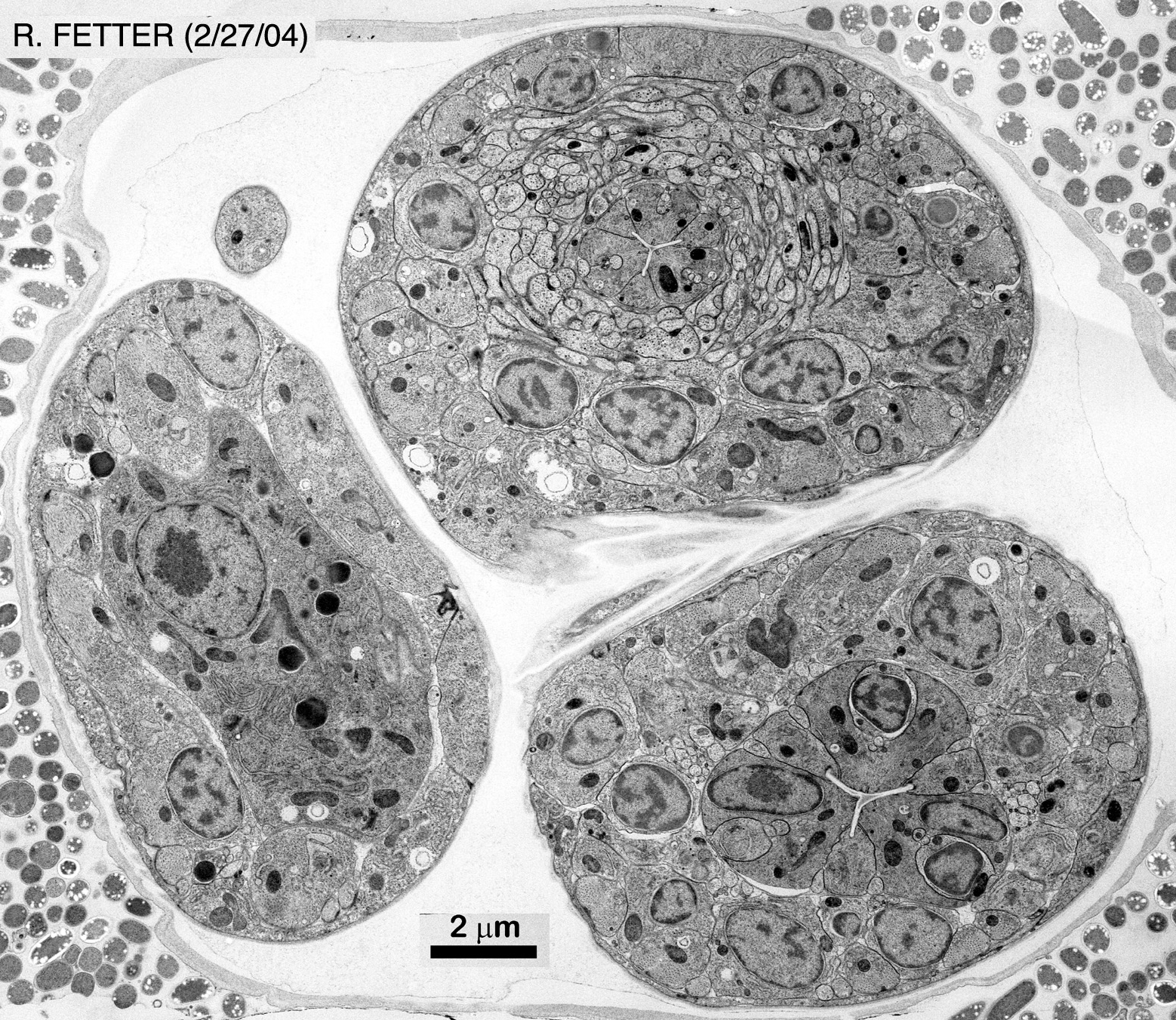
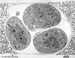
| 3-Fold Embryo (low magnification) |
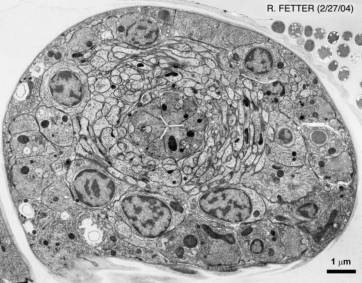
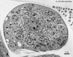
3-Fold Embryo (Nerve Ring) |
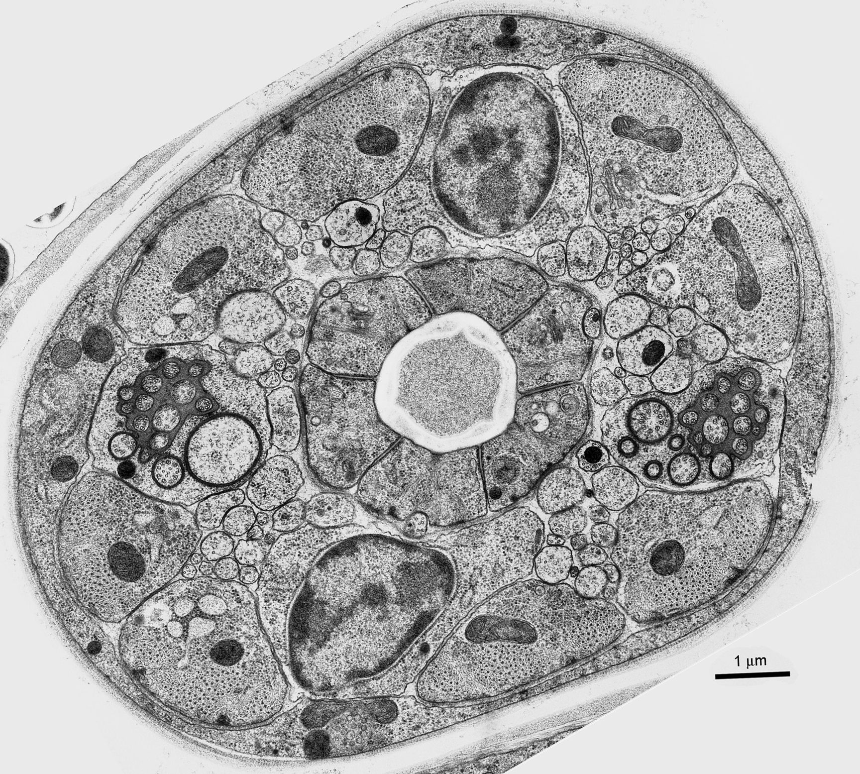
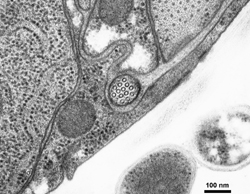
3-Fold Embryo (Amphid Cilia) |
 |
 |
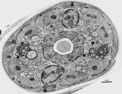 |
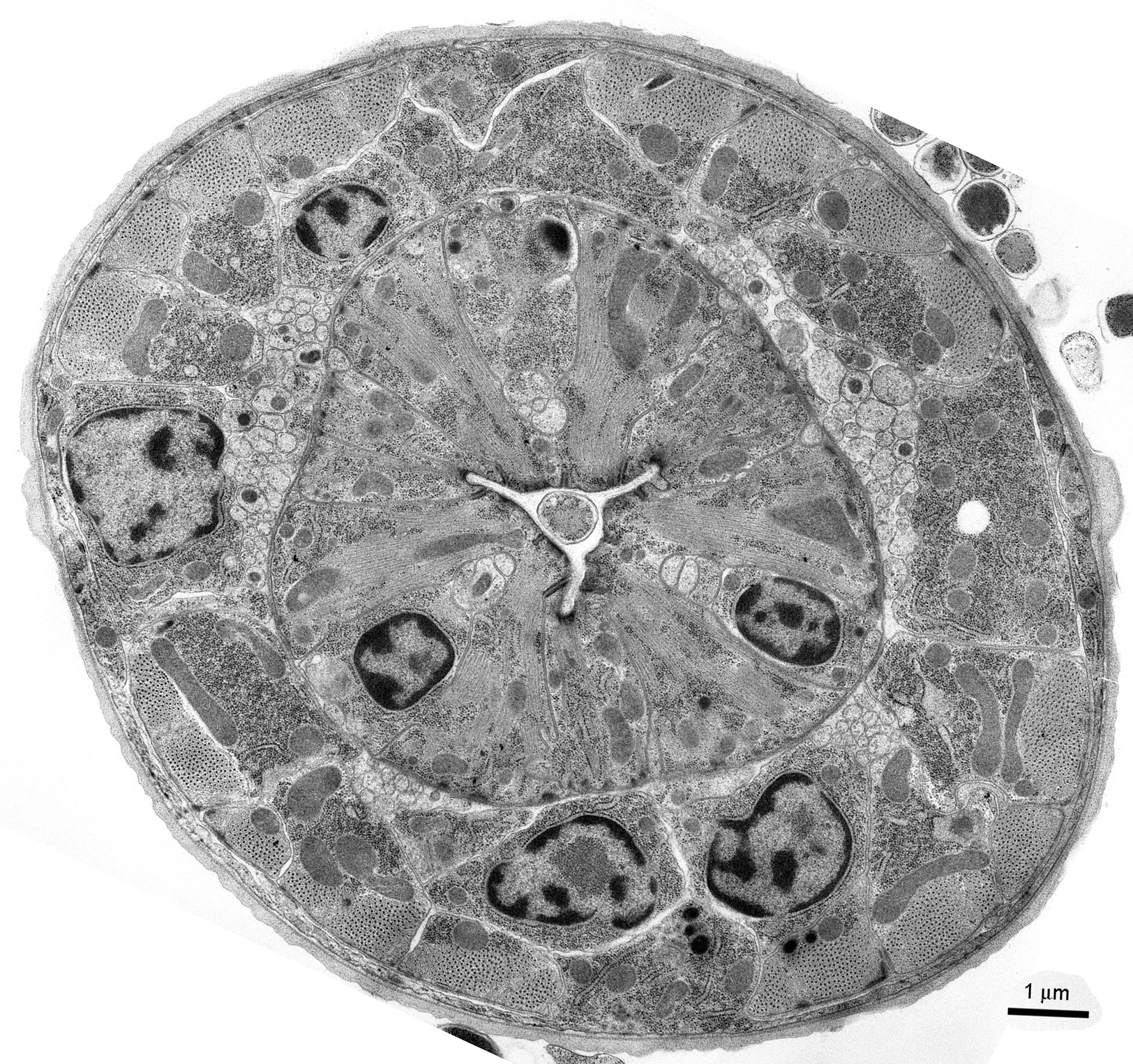
| L1 Larva (Pharynx) |
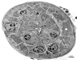
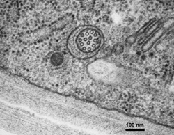
L1 Larva (ALM) |
L1 Larva (PLM) |
 |
 |
 |
Richard Fetter, Ph.D.
Senior Scientist
Janelia Farm Research Campus, HHMI
19700 Helix Drive
Ashburn, Virginia 20147
(Formerly) Dept. of Anatomy/HHMI
University of California, San Francisco
513 Parnassus Ave, Box 0452
San Francisco, CA 94143
References
Kennerdell, J.R., Fetter, R.D. and Bargmann, C.I. 2009. Wnt-Ror signaling to SIA and SIB neurons directs anterior axon guidance and nerve ring placement in C. elegans. Development 136: 3801-10. Article
Saalfeld, S., Fetter, R., Cardona, A. and Tomancak, P. 2012. Elastic volume reconstruction from series of ultra-thin microscopy sections. Nat. Methods 9: 717-20. Abstract
|