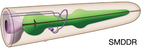
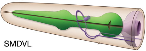
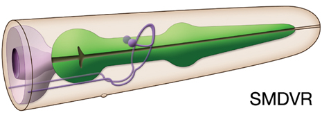
SMDDL, SMDDR, SMDVL, SMDVR

Type: Motor neuron
In MoW: SMD
Male Wiring Project:
SMDDL, SMDDR, SMDVL, SMDVR
In Wormbase: SMD, SMDDL, SMDDR, SMDVL, SMDVR
Lineage: AB plpapaaaa, AB prpapaaaa,
AB alppappaa, AB arappppaa
Location: The cell bodies of SMDV are located subdorsally in the head, posterior and close to the neuropile of the nerve ring.The cell bodies of SMDD are in the ventral ganglion
Description: These motor neurons innervate muscles in the head via NMJs in the nerve ring and also send processes posteriorly down the sublateral cords. 
Neurotransmitter/ Neuropeptide:
- Acetylcholine
(Loer, 2010; Duerr et al, 2008; Rand and Nonet, 1997-Appendix 2)
Innexin expression:
- UNC-7
- UNC-9
(Altun et al., 2009)
Receptor expression:
Function:
- Functions in locomotion: After animals are removed from bacterial food, they initiate a local search behavior consisting of reversals and deep omega-shaped turns. This is followed by dispersal ~30 min later as reversal and turns are suppressed. Local search behavior is triggered by AWC olfactory neurons, ASK gustatory neurons, and AIB interneurons while dispersal is promoted by ASI gustatory neurons and AIY interneurons (Gray et al., 2005). Downstream of AIB and AIY, motor neurons define specific aspects of reversal and turn frequency, amplitude, and directionality. SMD motor neurons define the steep amplitude of omega turns, RIV motor neurons specify the ventral bias of turns that follow a reversal, and SMB motor neurons set the amplitude of sinusoidal movement.


Click here for larger version
SMDDL
(AB plpapaaaa) development in the embryo.Dorsal view. Bottom is left side of the embryo. Spheres indicate individual nuclei. Black sphere: ancestors of SMDDL; dark grey spheres: apoptotic cells; other cells follow the WA color code (after they acquire specific cell or tissue identities). 0 min is fertilization. Click on the movie for higher resolution rendition (by A. Santella & Z. Bao).


Click here for larger version
SMDDR
(AB prpapaaaa) development in the embryo.Dorsal view. Bottom is left side of the embryo. Spheres indicate individual nuclei. Black sphere: ancestors of SMDDR; dark grey spheres: apoptotic cells; other cells follow the WA color code (after they acquire specific cell or tissue identities). 0 min is fertilization. Click on the movie for higher resolution rendition (by A. Santella & Z. Bao).


Click here for larger version
SMDVL (AB alppappaa) development in the embryo.
Dorsal view. Bottom is left side of the embryo. Spheres indicate individual nuclei. Black sphere: ancestors of SMDVL; dark grey spheres: apoptotic cells; other cells follow the WA color code (after they acquire specific cell or tissue identities). 0 min is fertilization. Click on the movie for higher resolution rendition (by A. Santella & Z. Bao).


Click here for larger version
SMDVR (AB arappppaa) development in the embryo.
Dorsal view. Bottom is left side of the embryo. Spheres indicate individual nuclei. Black sphere: ancestors of SMDVR; dark grey spheres: apoptotic cells; other cells follow the WA color code (after they acquire specific cell or tissue identities). 0 min is fertilization. Click on the movie for higher resolution rendition (by A. Santella & Z. Bao).


Last revision: July 31, 2014
image