|
|
|
 
|
PVQL, PVQR
Type: Interneuron
In MoW: PVQ
Male Wiring Project:
PVQL,
PVQR
In Wormbase: PVQ, PVQL, PVQR
Lineage: AB plapppaaa, AB prapppaaa
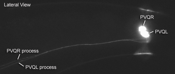
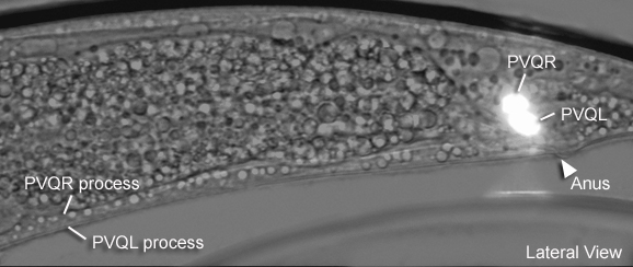
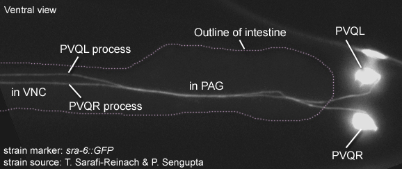
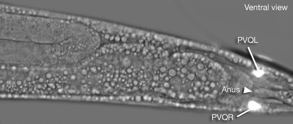
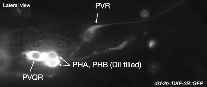
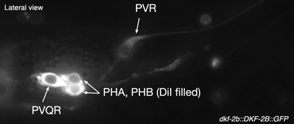
Location: Right and left lumbar ganglia
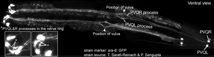
Description: Interneurons, project along ventral cord to ring. PVQL axon travels in the left tract while PVQR axon travels in the right track.
Neurotransmitter/ Neuropeptide:
- Glutamate
(Pereira et al., 2015)
Innexin expression:
- INX-3
- INX-7
- INX-13
- UNC-7
- UNC-9
(Altun et al., 2009)
Receptor expression:
- DOP-1; D1-like dopamine receptor
- Possibly GGR-1 ; GABA-A/glycine receptor-like protein
- GLR-1; glutamate receptor subunit
- GLR-5; glutamate receptor subunit
|


|
- SRA-6; G protein-coupled seven transmembrane receptor
- SER-1; serotonin receptor
- PDFR-1; pigment dispersing factor (PDF-1) receptor
(Wormbase; Barrios et al., 2012; Altun, 2011; Dernovici et al, 2007; Carnell et al, 2005; Tsalik et al., 2003; Brockie et al., 2001; Fujiwara et al., 1996; Troemel et al., 1995)
Function:
- Pioneering of the left tract of the ventral cord appears to be a joint action of PVPR and PVQL processes; PVQ axons pioneer the lumber commissures and continue to travel alongside the PVP processes in the VNC (Durbin, 1987; Wadsworth and Hedgecock, 1996; Wadsworth et al., 1996). When PVPR precursor is ablated in the embryo, PVQL and AVKR growth cones migrate towards the right side. (Durbin, 1987; Antebi et al., 1997)
|
 Click pictures for higher resolution images Click pictures for higher resolution images
 |
 |
 |
 |
 |
 |
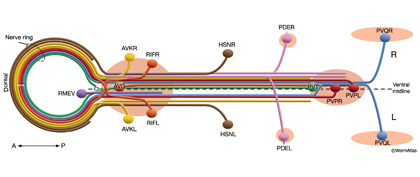
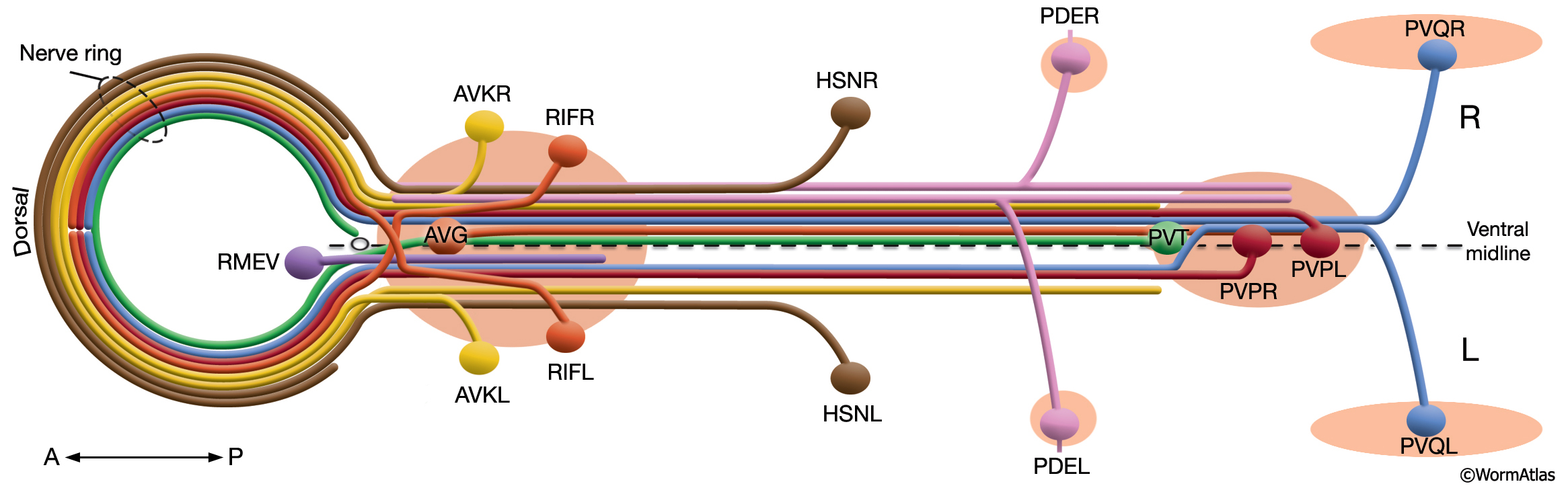
Schematic view of neurons that pioneer the left and right fascicles of the VNC. The body posterior to the NR is shown as if opened along the dorsal midline in cylindrical projection while the nerve ring is flattened toward the anterior. (Circle) Excretory pore at the anterior of the ventral midline; (light red ovals) ganglia. For clarity, neurons are given individual colors that are different from the color code used throughout this Atlas. (Based on White et al., 1986; Wadsworth et al., 1996.)
|
|
|
|
Click here for larger version
PVQL (AB plapppaaa) development in the embryo. Dorsal view. Bottom is left side of the embryo. Spheres indicate individual nuclei. Black sphere: ancestors of PVQL; dark grey spheres: apoptotic cells; other cells follow the WA color code (after they acquire specific cell or tissue identities). 0 min is fertilization. Click on the movie for higher resolution rendition (by A. Santella & Z. Bao). |
Click here for larger version
PVQR (AB prapppaaa) development in the embryo. Dorsal view. Bottom is left side of the embryo. Spheres indicate individual nuclei. Black sphere: ancestors of PVQR (since last PVQR ancestor has not yet gone through its final division, the black sphere seen at the end of this movie is still AB prapppaa); dark grey spheres: apoptotic cells; other cells follow the WA color code (after they acquire specific cell or tissue identities). 0 min is fertilization. Click on the movie for higher resolution rendition (by A. Santella & Z. Bao). |
|

Last revision: March 11, 2015 |