PDA

Type: Motor neuron
In MoW: PDA
Male Wiring Project:
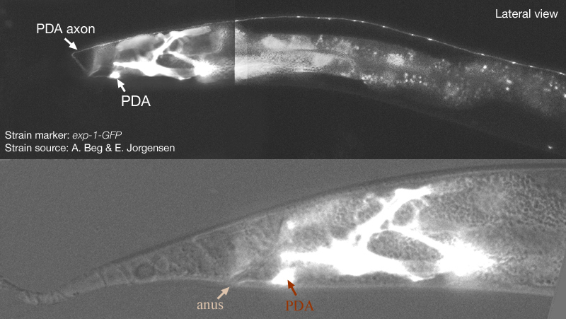
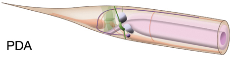
PDA
In Wormbase: PDA
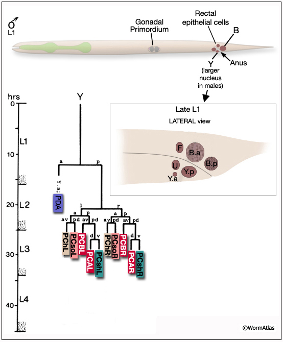
Lineage: AB prpppaaaa (in herm), Y.a (in male)
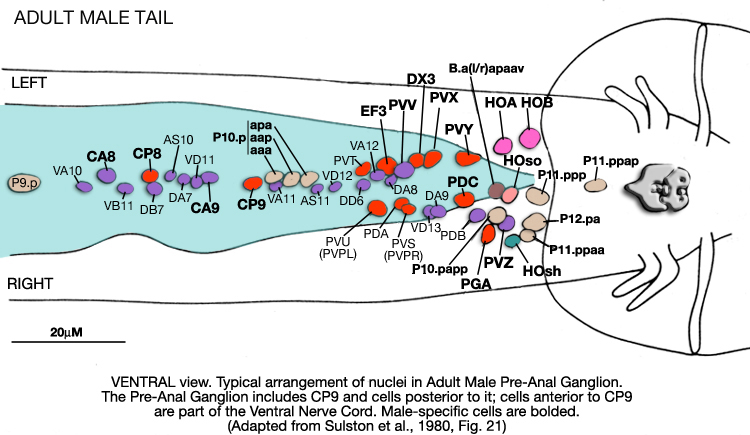
Location: Preanal ganglion
Description: During the second larval stage (L2), the rectal epithelial cell Y withdraws from the epithelium, migrates anterodorsally, and then becomes the PDA neuron through a transdifferentiation event while P12.pa, born at the end of the L1 stage just anterior to the position of Y, replaces Y in the rectum, completing the toroid with B epithelial cell. (Sulston and Horvitz 1977). The dorsal cord process of PDA ends in the posterior part of the body.


In male, Y divides and the anterior daughter becomes PDA. Hermaphrodites and males exhibit some differences in PDA connectivity.
Neurotransmitter/ Neuropeptide:
- Acetylcholine
(Pereira et al., 2015)
Innexin expression:
- UNC-9
(Altun et al., 2009)
Receptor expression:
- EXP-1; excitatory GABA receptor subunit
- UNC-8, a DEG/ENaC family member homologous to subunits of a candidate mechanically gated ion channel
(Beg and Jorgensen, 2003, also pers. comm.; Tavernarakis et al., 1997)
Function:
- Innervates posterior dorsal body wall muscles


Click image for closeup view Click pictures for higher resolution images

HERMAPHRODITE


MALE


Click here for larger version
PDA (AB prpppaaaa) development in the embryo.
Dorsal view. Bottom is left side of the embryo. Spheres indicate individual nuclei. Black sphere: ancestors of PDA; dark grey spheres: apoptotic cells; other cells follow the WA color code (after they acquire specific cell or tissue identities). 0 min is fertilization. Click on the movie for higher resolution rendition (by A. Santella & Z. Bao).


Last revision: April1, 2014
image