Click here for larger version
OLLL (AB alppppapaa) development in the embryo. Dorsal view. Bottom is left side of the embryo. Spheres indicate individual nuclei. Black sphere: ancestors of OLLL (since last OLLL ancestor has not yet gone through its final division, the black sphere seen at the end of this movie is still AB alppppapa); dark grey spheres: apoptotic cells; other cells follow the WA color code (after they acquire specific cell or tissue identities). 0 min is fertilization. Click on the movie for higher resolution rendition (by A. Santella & Z. Bao).
Click here for larger version
OLLR (AB praaapapaa) development in the embryo. Dorsal view. Bottom is left side of the embryo. Spheres indicate individual nuclei. Black sphere: ancestors of OLLR (since last OLLR ancestor has not yet gone through its final division, the black sphere seen at the end of this movie is still AB paaapapa); dark grey spheres: apoptotic cells; other cells follow the WA color code (after they acquire specific cell or tissue identities). 0 min is fertilization. Click on the movie for higher resolution rendition (by A. Santella & Z. Bao).
OLL Sensory Endings
Click here for larger version
3D reconstruction of the anterior sensory endings (cilia and dendrites) from high resolution serial section transmission electron micrographs. Bar 1 μm. Color code for the sensory endings is shown on the right-colors do not follow the WA color code. To expand, double click on the video, to return to original size, click "esc" (Doroquez et al., 2014)
Click here for larger version
3D reconstruction of all amphid neuron cilia and associated socket and sheath cell processes. Modeled from serial section transmission electron micrographs (ssTEMs). Bar 1 μm. Color code for the sensory endings is shown on the left-colors do not follow the WA color code. To expand, double click on the video, to return to original size, click "esc" (Doroquez et al., 2014)
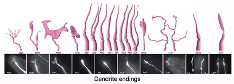
Click here for larger version
3D reconstruction of IL1, IL2, OLL, OLQ and CEP sensory endings and associated glia. Colors do not follow the WA color code (purple; transition zone (TZ), lavender; CEP, blue; OLL and OLQ, beige; IL1, green; IL2, light pink; ILsh, dark pink; ILso, dark orange; OLso, light orange; OLsh, dark green; CEPso, light green; CEPsh).To expand, double click on the video, to return to original size, click "esc" (Doroquez et al., 2014)


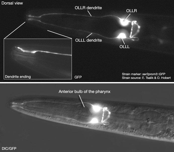
 Click pictures for higher resolution images
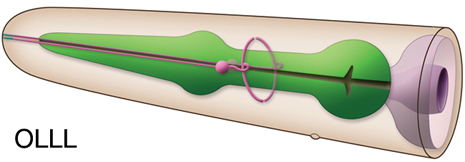
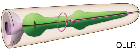