to the metacorpus and possibly the isthmus. They are bipolar neurons with a posterior dendrite and an anterodorsal axon. The posterior process is approximately 5 µm and makes an occasional single synapse to pm5. The anterior projection of these cells grows past the nerve ring, and sends one branch to the dorsal side through pm4 similar to the process of M2 neuron. An additional branch runs posteriorly between pm4 and pm5 at the outside of the pharynx to just behind the nerve ring and terminates with neuromuscular synapses on the posterior edge of pm4. The branches to the dorsal side make no neuromuscular synapses along their dorsal trajectory, but similar to the two subventral branches, they pass posteriorly between pm4 and pm5 at the dorsal edge of the pharynx and form synapses on pm4 behind the nerve ring (Albertson and Thomson, 1976)
Neurotransmitter/ Neuropeptide:
- Glutamate
- FLP-18 ; FMRFamide-related neuropeptide (FaRP)
- NLP-3; neuropeptide-like protein
(Rogers et al., 2003; Nathoo et al., 2001; Lee et al., 1999; Radice and Visser, 1996)
Innexin expression:
- INX-3
(Altun et al., 2009)
Receptor expression:
- GLR-8; glutamate receptor subunit
- NPR-1; neuropeptide Y receptor like protein
(Coates and de Bono, 2002; Brockie et al., 2001)
Function:
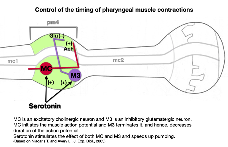
- M3s modulate the timing of pharyngeal relaxation in corpus and promote rapid relaxation of the pharyngeal muscle following contraction. When M3s are decoupled from the pharyngeal muscle by mutating the postsynaptic receptor (a glutamate-gated chloride channel partly encoded by avr-15) for M3 neurotransmission, the duration of pharyngeal contraction increases (Dent et al., 1997). Each M3 is suggested to be proprioceptive and fire in response to corpus muscle contraction, which, in turn, induces inhibitory postsynaptic potentials (repolarization) and relaxation of the muscle. This relaxation seems to be important for effective transport of bacteria within the lumen (Avery and Thomas, 1997). Exogenous serotonin mimics the presence of food and induces rapid contraction-relaxation cycles with shortened action potentials in pharynx. This effect is dependent on MC and M3 neurons (see model below) (Niacaris and Avery, 2003) |


 Click pictures for higher resolution images