M1

Type: Pharyngeal motor neuron
In MoW:
Male Wiring Project: M1
In Wormbase: M1
Lineage: MS paapaaa
Location: Pharynx posterior bulb
Description: The cell body of M1 is located subdorsally between the muscle and right marginal cell. A single process runs anteriorly in this position to the nerve ring where it then joins the dorsal nerve cord. Upon reaching the level of


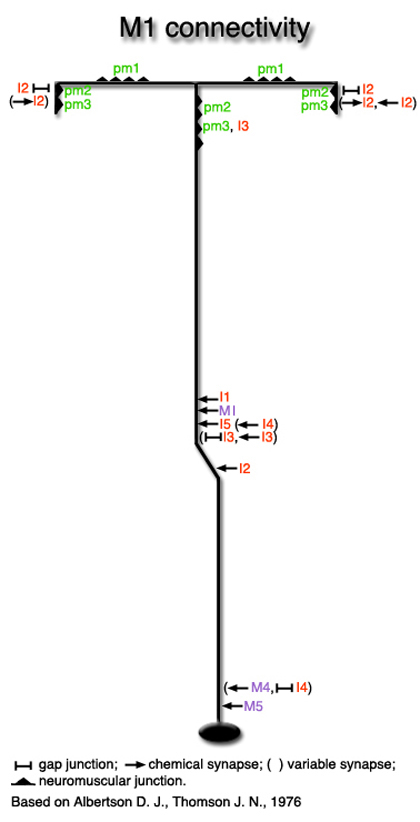
pm1, the process splits into two to send a vesicle-filled process to each of the subventral nerve cords, synapsing on pm1 and pm2 before reaching the cords. These then run posteriorly for a short length and form synapses on pm2 and pm3 (Albertson and Thomson, 1976)
Neurotransmitter/ Neuropeptide:
- Acetylcholine
- NLP-3 ; neuropeptide-like protein
(Loer, 2010; Duerr et al, 2008; Nathoo et al., 2001)
Innexin expression:
- INX-5
- INX-13
(Altun et al., 2009)
Receptor expression:
- GLR-2; ionotropic glutamate receptor subunit
- Possibly AVR-14; a subunit of the glutamate-gated chloride channel
(Cook et al., 2006; Brockie and Maricq, 2006; Brockie et al., 2001).
Function: Unknown or redundant (Avery and Thomas, 1997)


Click image for closeup view Click pictures for higher resolution images

Variable synapses to and from I2 neuron shown here are not included in the I2 neuron connectivity diagram

See also Pharynx Atlas


Click here for larger version
M1 (MS paapaaa) development in the embryo.
Dorsal view. Bottom is left side of the embryo. Spheres indicate individual nuclei. Black sphere: ancestors of M1; dark grey spheres: apoptotic cells; other cells follow the WA color code (after they acquire specific cell or tissue identities). 0 min is fertilization. Click on the movie for higher resolution rendition (by A. Santella & Z. Bao).


Last revision: February 4, 2014
image