|
|
|
 
|
I1L, I1R
Type: Pharyngeal interneuron (also anterior sensory?)
In MoW:
Male Wiring Project: I1L, I1R
In Wormbase: I1, I1L, I1R
Lineage: AB alpapppaa, AB arapappaa
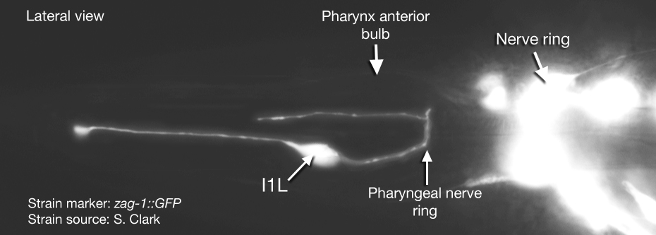
Location: Pharynx anterior bulb
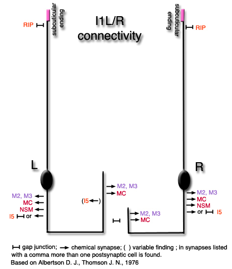
Description: Pharyngeal interneurons. The anterior projections from these cells insert onto muscles pm1 and pm2s, and have a free subcuticular, proprioceptive-like (postulated sensory) ending at the anterior end. They also extend to the basement membrane here to receive input from the somatic RIP neurons. The posterior axonal projections of the I1s run within the subventral nerve cords and then run around the pharyngeal nerve ring to the dorsal side where they meet in gap junctions. The termination of the |
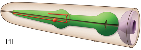
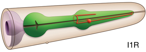
|
anterior projections in the dorsal pharyngeal nerve cord is variable (Albertson and Thomson, 1976)
Neurotransmitter/Neuropeptide:
- Acetylcholine
- NLP-3 ; neuropeptide-like protein
(Pereira et al., 2015; Loer, 2010; Duerr et al, 2008; Nathoo et al., 2001)
Innexin expression:
- UNC-9
(Altun et al., 2009)
Receptor expression:
- GLR-7; glutamate receptor subunit
- GLR-8; glutamate receptor subunit
(Brockie et al., 2001)
Function:
- The pharyngeal and somatic nervous systems are connected to each other via gap junctions between the extrapharyngeal RIP neurons and the pharyngeal I1 neurons. When this connection is disrupted by ablation of RIPs, pharyngeal pumping becomes unresponsive to light touch which is sensed by somatic touch neurons (Avery and Thomas, 1997). I1s also synapse on MC neurons and modulate the rate of pharyngeal pumping in the absence of bacteria (Avery and Thomas, 1997) |
 Click pictures for higher resolution images Click pictures for higher resolution images
|
|
Click here for larger version
I1L (AB alpapppaa) development in the embryo. Dorsal view. Bottom is left side of the embryo. Spheres indicate individual nuclei. Black sphere: ancestors of I1L; dark grey spheres: apoptotic cells; other cells follow the WA color code (after they acquire specific cell or tissue identities). 0 min is fertilization. Click on the movie for higher resolution rendition (by A. Santella & Z. Bao). |
Click here for larger version
I1R (AB arapappaa) development in the embryo. Dorsal view. Bottom is left side of the embryo. Spheres indicate individual nuclei. Black sphere: ancestors of I1R; dark grey spheres: apoptotic cells; other cells follow the WA color code (after they acquire specific cell or tissue identities). 0 min is fertilization. Click on the movie for higher resolution rendition (by A. Santella & Z. Bao). |
|

Last revision: DEcember 17, 2013 |