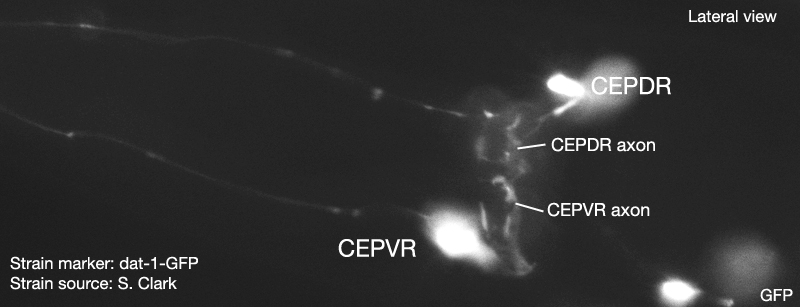
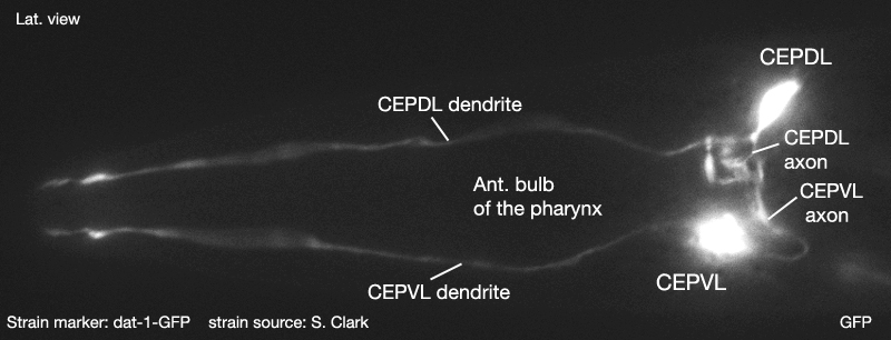
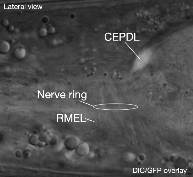
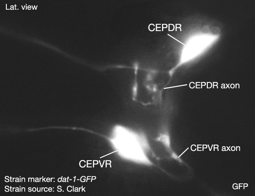
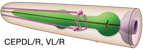
Type: Sensory neuron (mechanosensory) In MoW:CEP Male Wiring Project:CEPDL,
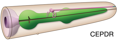
CEPDR,
CEPVL,
CEPVR In Wormbase:CEP, CEPDL, CEPDR, CEPVL, CEPVR Lineage:AB plaaaaappa, AB arpapaappa
AB plpaappppa, AB prpaappppa Location: Head Description:Neurons of cephalic sensilla. CEP dendrite extends to the tip of the nose within corresponding labial nerve. CEPD neurons are directly exposed to the pseudocoelomic body fluid. CEP express acetylcholinesterase of class A, ACE-1(Culetto et al., 1999) Neurotransmitter/ Neuropeptide:
- Dopamine (Lints and Emmons, 1999) Innexin expression:
- UNC-9 (Altun et al., 2009) Receptor expression:
- DOP-2; dopamine receptor (Suo et al., 2003) Function:
- Mechanosensensation in the head; deflection of its single cilium by pressure application produces ion currents in CEP neuron, consistent with mechanosensory function (Kang et al., 2010). The three classes of dopaminergic neurons (CEP, ADE, and PDE) function redundantly to sense the mechanosensory stimulus from bacteria and mediate the motor circuit to control "basal slowing response"; when well-fed C. elegans hermaphrodites are washed clean of bacteria and then reintroduced to the bacterial lawn, they move more slowly than when transferred to an environment without bacteria. (Sawin et al., 2000)
Click pictures for higher resolution images
Embryonic development of CEP neurons
Click here for larger version
CEPDL (AB plaaaaappa) development in the embryo. Dorsal view. Bottom is left side of the embryo. Spheres indicate individual nuclei. Black sphere: ancestors of CEPDL; dark grey spheres: apoptotic cells; other cells follow the WA color code (after they acquire specific cell or tissue identities). 0 min is fertilization. Click on the movie for higher resolution rendition (by A. Santella & Z. Bao).
Click here for larger version
CEPDR (AB arpapaappa) development in the embryo. Dorsal view. Bottom is left side of the embryo. Spheres indicate individual nuclei. Black sphere: ancestors of CEPDR; dark grey spheres: apoptotic cells; other cells follow the WA color code (after they acquire specific cell or tissue identities). 0 min is fertilization. Click on the movie for higher resolution rendition (by A. Santella & Z. Bao).
Click here for larger version
CEPVL (AB plpaappppa) development in the embryo. Dorsal view. Bottom is left side of the embryo. Spheres indicate individual nuclei. Black sphere: ancestors of CEPVL; dark grey spheres: apoptotic cells; other cells follow the WA color code (after they acquire specific cell or tissue identities). 0 min is fertilization. Click on the movie for higher resolution rendition (by A. Santella & Z. Bao).
Click here for larger version
CEPVR (AB prpaappppa) development in the embryo. Dorsal view. Bottom is left side of the embryo. Spheres indicate individual nuclei. Black sphere: ancestors of CEPVR; dark grey spheres: apoptotic cells; other cells follow the WA color code (after they acquire specific cell or tissue identities). 0 min is fertilization. Click on the movie for higher resolution rendition (by A. Santella & Z. Bao).
CEP Sensory Endings
Click here for larger version
3D reconstruction of the anterior sensory endings (cilia and dendrites) from high resolution serial section transmission electron micrographs. Bar 1 μm. Color code for the sensory endings is shown on the right-colors do not follow the WA color code. To expand, double click on the video, to return to original size, click "esc" (Doroquez et al., 2014)
Click here for larger version
3D reconstruction of all amphid neuron cilia and associated socket and sheath cell processes. Modeled from serial section transmission electron micrographs (ssTEMs). Bar 1 μm. Color code for the sensory endings is shown on the left-colors do not follow the WA color code. To expand, double click on the video, to return to original size, click "esc" (Doroquez et al., 2014)
Click here for larger version
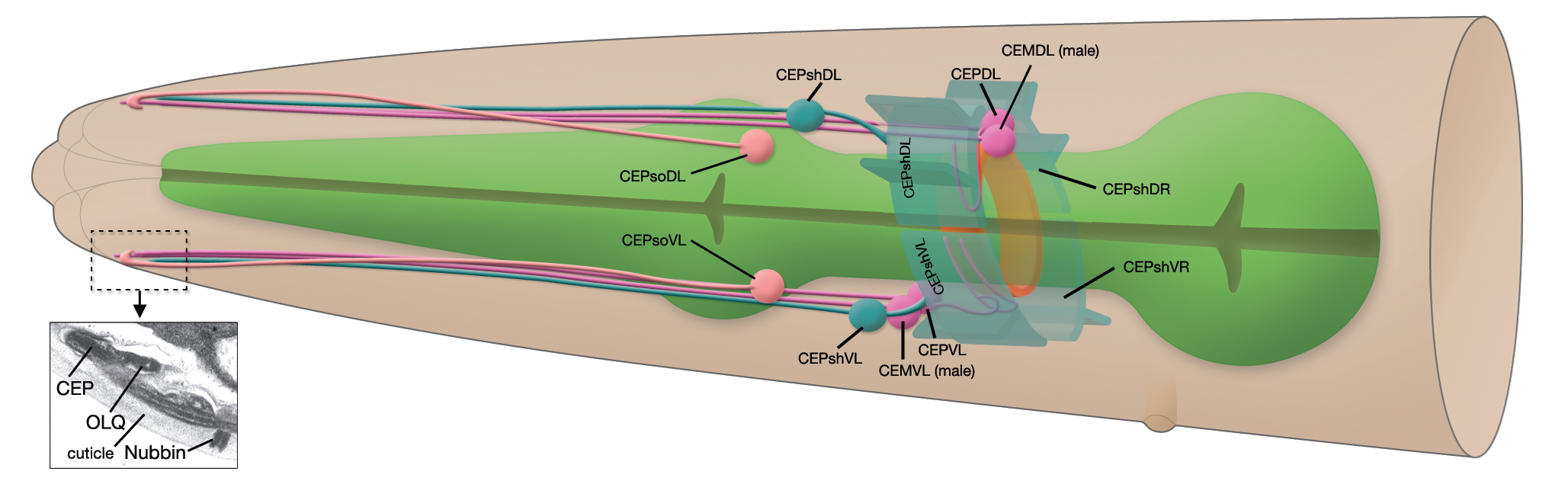
3D reconstruction of IL1, IL2, OLL, OLQ and CEP sensory endings and associated glia. Colors do not follow the WA color code (purple; transition zone (TZ), lavender; CEP, blue; OLL and OLQ, beige; IL1, green; IL2, light pink; ILsh, dark pink; ILso, dark orange; OLso, light orange; OLsh, dark green; CEPso, light green; CEPsh).To expand, double click on the video, to return to original size, click "esc" (Doroquez et al., 2014)


 Click pictures for higher resolution images