Type: Sensory neuron (chemosensory (gustatory), thermosensory) In MoW:ASI Male Wiring Project:ASILh,
ASIRh,
ASILm,
ASIRm In Wormbase:ASI, ASIL, ASIR Lineage: AB plaapapppa, AB praapapppa Location: Lateral ganglia of head Description:Amphid neurons, single (AsI) ciliated endings Like all other amphid neurons, ASI are born near the presumptive nose of the embryo during development. They then anchor a short projection there, after which the cell body migrates away, stretching the dendrite out behind it. This process is dependent on DEX-1 or DYF-7, secreted extracellular matrix proteins which act cooperatively for anchoring. In mutants lacking these proteins, the dendrite fails to anchor at the nose and is dragged along with the migrating cell body, giving rise to a short dendritic stub (Heiman and Shaham, 2010). Dendritic process takes up FITC. ASI axon projects into the ventral cord by way of the same side amphid commissure and then grows into the nerve ring where it makes diverse synaptic connections in ring neuropil. Neurotransmitter/Neuropeptide:
- DAF-28; beta-type insulin, homologous to human insulin
- FLP-2; FMRFamide-like peptide; variably expressed/neurons tentatively identified
- FLP-10; FMRFamide-like peptide
- FLP-21; FMRFamide-like peptide
- INS-1; insulin-like peptide, orthologous
to human insulin
- INS-3; insulin-like peptide
- INS-4; insulin-like peptide
- INS-6; beta-type insulin-like molecule
- INS-7; insulin-like peptide
- INS-9; insulin-like peptide
- INS-22; insulin-like molecule, lacks a C-peptide domain
- INS-26; insulin-like peptide
- INS-32; insulin-like peptide
- NLP-1; neuropeptide-like protein
- NLP-5; neuropeptide-like protein
- NLP-6; neuropeptide-like protein
- NLP-7; neuropeptide-like protein - NLP-9; neuropeptide-like protein
- NLP-14; neuropeptide-like protein
- NLP-18; neuropeptide-like protein
- NLP-24; neuropeptide-like protein
- NLP-27; neuropeptide-like protein
- PDF-1; pigment dispensing factor homolog (Pierce et al., 2013; Cornils et al., 2011; Janssen et al., 2009; Takayama et al., 2009; Li and Kim, 2008; Kodama et al., 2006; Tomioka et al., 2006; Li et al., 2003; Nathoo et al., 2001; Pierce et al., 2001) Innexin expression:
- INX-18
- INX-19 (Altun et al., 2009; Chuang et al., 2007) Receptor Expression:
- DAF-11; transmembrane receptor type guanylate cyclase
- DAF-37; a G protein-coupled receptor, which, cooperatively with DAF-38, mediates ascaroside perception
- DAF-38; a G protein-coupled receptor, which, cooperatively with DAF-37, mediates ascaroside perception
- DCAR-1; seven transmembrane receptor for dihydrocaffeic acid (a water-soluble repellent)
- GCY-1; transmembrane receptor type guanylate cyclase
- GCY-2; transmembrane receptor type guanylate cyclase
- GCY-3; transmembrane receptor type guanylate cyclase
- GCY-27; receptor type guanylate cyclase, lacks transmembrane domain and predicted signal sequence
- NPR-5; receptor for flp-3 and flp-18 encoded peptides
- NPR-15; neuropeptide receptor
- SER-6; octopamine receptor
- SRD-1; G protein-coupled seven transmembrane receptor
- STR-2; G protein-coupled seven transmembrane receptor
- STR-3; G protein-coupled seven transmembrane receptor (Wormbase; Mills et al., 2012; Park et al., 2012; Altun, 2011; Aoki et al., 2011; Ortiz et al., 2006; Peckol et al.. 2001; Birnby et al., 2000) Function:The main synpatic output of ASI is onto AIA interneurons. The ASI neurons modulate behavior in several contexts, so they may work to integrate information about the environment (such as population density, food availability, CO2 level or the presence of sex pheromone) and adjust either the function or programming of related neural circuits via DAF-7/TGF-β (White and Jorgensen, 2012.) ASI neurons are the sole source of DAF-7/TGF-β in C. elegans.
- Function in locomotion: Suppress omega turns and reversals enhancing dispersal (Gray et al., 2005). After animals are removed from bacterial food, they initiate a local search behavior consisting of reversals and deep omega-shaped turns. This is followed by dispersal ~30 min later as reversal and turns are suppressed. Local search behavior is triggered by AWC olfactory neurons, ASK gustatory neurons, and AIB interneurons while dispersal is promoted by ASI gustatory neurons and AIY interneurons (Gray et al., 2005).
- Chemotaxis: ASI neurons are involved in chemotaxis to lysine, contribute to a residual chemotactic response to cAMP, biotin, Cl-, and Na+ after ASE neurons are killed (Bargmann and Horvitz, 1991)
- Sense temperature: After cultivation at a uniform temperature (Tc) with sufficient food, animals preferentially migrate to their cultivation temperature (Tc) when placed on a thermal gradient, and move isothermally at this temperature (Hedgecock and Russell 1975.) Animals sense and record their Tc by AFD (major thermosensory), AWC and ASI neurons (Beverly et al, 2011; Kuhara et al., 2008; Biron et al., 2008.) This Tc memory is plastic and can be reset upon cultivation at a different temperature (Hedgecock and Russell 1975.) Unlike AFD neurons, which respond to thermal stimuli above Tc with continuous, graded calcium signals in a deterministic and highly reproducible way, ASI neurons exhibit temperature-induced stochastic Ca++ transients in a defined operating range with decreased responses close to or far from Tc (Beverly et al, 2011)
- Sense pheromone and are involved in behavioral responses to pheromone (C. elegans pheromone is a mixture composed of derivatives of the dideoxysugar ascarylose (C3, C6, C7, C9 and other ascarosides) that regulates entry to dauer stage at L1, as well as adult behaviors such as sexual attraction of males to hermaphrodites and social behavior, i.e., attraction between hermaphrodites to clump into feeding groups)(Park et al., 2012.) ADF, ASI and ASG inhibit entry into dauer stage while ASJ and ASK promote dauer entry (Kim et al., 2009; Ouellett et al., 2008; Schackwitz et al., 1996; Bargmann and Horvitz, 1991.) ASI neurons function to repress pheromone actions (e.g, sexual attraction) in hermaphrodites; daf-7mutation or ablation of the ASI neuron pair reveals sexual attraction (male-specific behavior) in hermaphrodites (White and Jorgensen, 2012.) In turn, pheromone induces dauer formation in part by repression of DAF-7/TGF-β expression in ASI
- Modulate innate immune response to pathogenic bacteria. ASH, ASI, AQR, PQR, and URX neurons control the unfolded protein response (UPR) and a p38 mitogen-activated protein kinase signaling pathway required for innate immunity (Singh & Aballay, 2012.) Loss of OCTR-1 signaling in ASH and ASI increases the general immune function of the nematodes; OCTR-1 functions in these neurons to actively suppress innate immune responses by down-regulating the expression of noncanonical UPR genes in nonneuronal tissues (Sun et al., 2011)
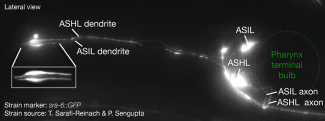
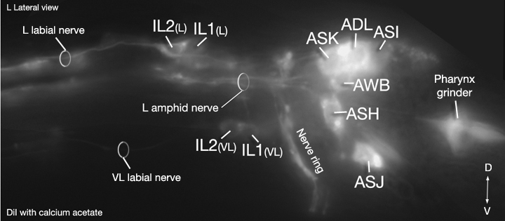
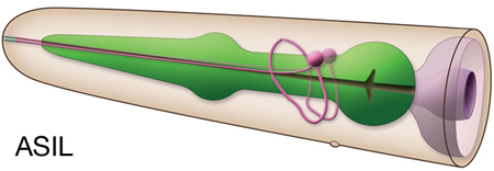
Click pictures for higher resolution images. Also see DiI staining of ASI in dorso-ventral view
Click here for larger version
ASIL (AB plaapapppa) development in the embryo. Dorsal view. Bottom is left side of the embryo. Spheres indicate individual nuclei. Black sphere: ancestors of ASIL (since last ASIL ancestor has not yet gone through its final division, the black sphere seen at the end of this movie is still AB plaapappp); dark grey spheres: apoptotic cells; other cells follow the WA color code (after they acquire specific cell or tissue identities). 0 min is fertilization. Click on the movie for higher resolution rendition (by A. Santella & Z. Bao).
Click here for larger version
ASIR (AB praapapppa) development in the embryo. Dorsal view. Bottom is left side of the embryo. Spheres indicate individual nuclei. Black sphere: ancestors of ASIR (since last ASIR ancestor has not yet gone through its final division, the black sphere seen at the end of this movie is still AB praapappp); dark grey spheres: apoptotic cells; other cells follow the WA color code (after they acquire specific cell or tissue identities). 0 min is fertilization. Click on the movie for higher resolution rendition (by A. Santella & Z. Bao).
Click here for larger version
3D reconstruction of the anterior sensory endings (cilia and dendrites) from high resolution serial section transmission electron micrographs (ssTEMs). Bar 1 μm. Color code for the sensory endings is shown on the right-colors do not follow the WA color code. To expand, double click on the video, to return to original size, click "esc" (Doroquez et al., 2014)
Click here for larger version
3D reconstruction of all amphid neuron cilia and associated socket and sheath cell processes. Modeled from serial section transmission electron micrographs (ssTEMs). Bar 1 μm. Color code for the sensory endings is shown on the left-colors do not follow the WA color code. To expand, double click on the video, to return to original size, click "esc" (Doroquez et al., 2014)


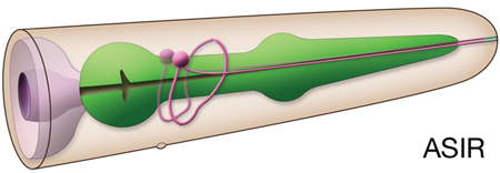

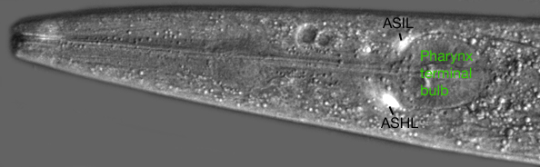
 Click pictures for higher resolution images. Also
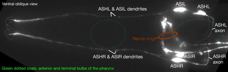