Type: Sensory neuron (chemosensory (gustatory)) In MoW:ASG Male Wiring Project:ASGLh,
ASGRh,
ASGLm,
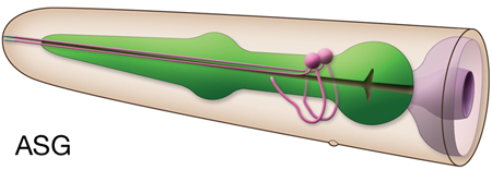
ASGRm In Wormbase: ASG, ASGL, ASGR Lineage:AB plaapapap, AB praapapap Location:Lateral ganglia of head Description: Amphid neurons, single (AsG) ciliated endings, Like all other amphid neurons, ASG are born near the presumptive nose of the embryo during development. They then anchor a short projection there, after which the cell body migrates away, stretching the dendrite out behind it. This process is dependent on DEX-1 or DYF-7, secreted extracellular matrix proteins which act cooperatively for anchoring. In mutants lacking these proteins, the dendrite fails to anchor at the nose and is dragged along with the migrating cell body, giving rise to a short dendritic stub (Heiman and Shaham, 2010). ASG axon projects into ring via amphid commissure from ventral ganglion and makes diverse synaptic connections in ring neuropil.
Developmental default state of the ASG neurons is AWA-like, and this fate must be repressed in order for the ASG neurons to adopt ASG fate. ALR-1 acts in parallel to UNC-130 and upstream of LIN-11 to specify the AWA and ASG neurons. (Melkman and Sengupta; Sarafi-Reinach and Sengupta, 2000) Neurotransmitter/ Neuropeptide:
- Glutamate
- Serotonin (under hypoxic conditions)
- FLP-6; FMRFamide-like neuropeptide
- FLP-13; FMRFamide-like neuropeptide
- FLP-22; FMRFamide-like neuropeptide
Click here for larger version
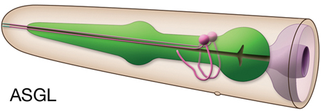
ASGL (AB plaapapap) development in the embryo. Dorsal view. Bottom is left side of the embryo. Spheres indicate individual nuclei. Black sphere: ancestors of ASGL; dark grey spheres: apoptotic cells; other cells follow the WA color code (after they acquire specific cell or tissue identities). 0 min is fertilization. Click on the movie for higher resolution rendition (by A. Santella & Z. Bao).
Click here for larger version
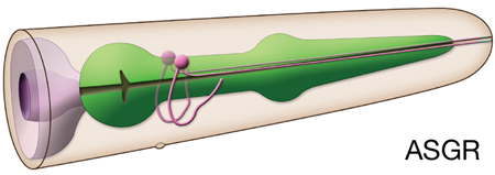
ASGR (AB praapapap) development in the embryo. Dorsal view. Bottom is left side of the embryo. Spheres indicate individual nuclei. Black sphere: ancestors of ASGR; dark grey spheres: apoptotic cells; other cells follow the WA color code (after they acquire specific cell or tissue identities). 0 min is fertilization. Click on the movie for higher resolution rendition (by A. Santella & Z. Bao).
Click here for larger version
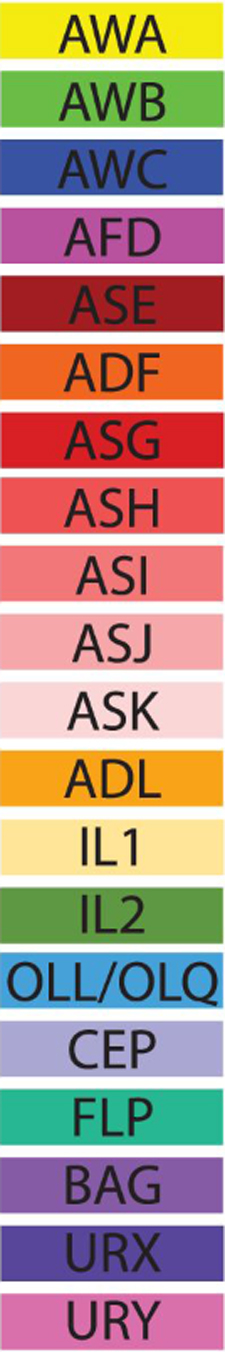
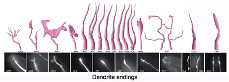
3D reconstruction of the anterior sensory endings (cilia and dendrites) from high resolution serial section transmission electron micrographs (ssTEMs). Bar 1 μm. Color code for the sensory endings is shown on the right-colors do not follow the WA color code. To expand, double click on the video, to return to original size, click "esc" (Doroquez et al., 2014)
Click here for larger version
3D reconstruction of all amphid neuron cilia and associated socket and sheath cell processes. Modeled from serial section transmission electron micrographs (ssTEMs). Bar 1 μm. Color code for the sensory endings is shown on the left-colors do not follow the WA color code. To expand, double click on the video, to return to original size, click "esc" (Doroquez et al., 2014)


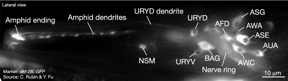
 Click pictures for higher resolution images
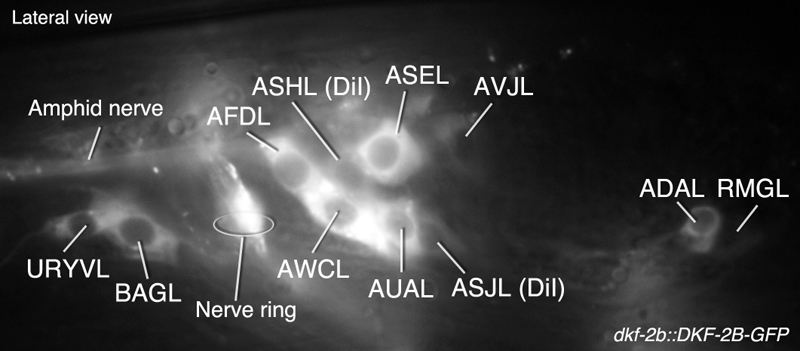