|
|
|
 
|
ALA
Type: Interneuron, mechanosensory neuron
In MoW: ALA
In WormWiring:
ALAh,
ALAm,
In Wormbase: ALA
Lineage: AB alapppaaa
Location: Dorsal ganglion of the head
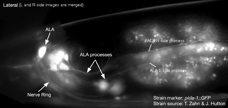
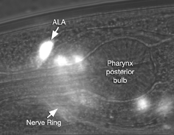
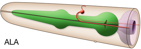
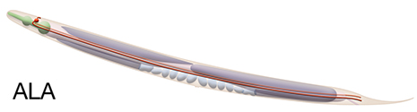
Description: ALA neuron has two processes that branch from the anterior portion of the cell body and project into the left and right sides of the nerve ring. A third, short process enters the dorsal cord and then peters out. The two large processes run on the left and rights sides of the ring and then leave the ring to join the lateral cords. They extend as far as the tail, adjacent to the excretory canals
Neurotransmitter/ Neuropeptide:
- FLP-7; FMRFamide-like peptide
(Li and Kim, 2008)
Innexin expression:
None yet reported, although described to have gap junctions in adult animals (MoW) |
|
Receptor expression:
- ACR-13 (LEV-8)
- ACR-4 (DES-2)
- SRA-10
(Wormbase; Altun, 2011; Van Buskirk and Sternberg, 2010; Towers et al., 2005; Troemel et al., 1995)
Function:
- Involved in inducing normal lethargus quiescence (i.e., cessation of pharyngeal pumping and locomotion during the lethargus periods (an EGF/LET-23-induced sleep-like state prior to molts). ALA neuron inhibits locomotion by inhibiting AVE, which normally functions to promote locomotion. Synapse between ALA and AVE is contacted by the CEPsh glia and CEPsh cells inhibit synaptic transmission from ALA to AVE, promoting locomotion.
(Mulcahy and Ient 2010; Van Buskirk and Sternberg, 2007)
- ALA is a high-threshold mechanosensor, which responds to harsh, e.g.with a pick, mechanical stimuli and is involved in inhibition of egg-laying in response to these stimuli. It doesn't respond to light touch. Also, its physiological responses to anterior vs posterior touch are distinct suggesting it can distinguish between spacially separated stimuli. Its laterally placed long processes are required for this harsh-touch response.
(Sanders et al., 2013)
|
 Click pictures for higher resolution images Click pictures for higher resolution images
|
|
Click here for larger version
ALA (AB alapppaaa) development in the embryo. Dorsal view. Bottom is left side of the embryo. Spheres indicate individual nuclei. Black sphere: ancestors of ALA; dark grey spheres: apoptotic cells; other cells follow the WA color code (after they acquire specific cell or tissue identities). 0 min is fertilization. Click on the movie for higher resolution rendition (by A. Santella & Z. Bao). |
|
|

Last revision: November 26, 2013 |