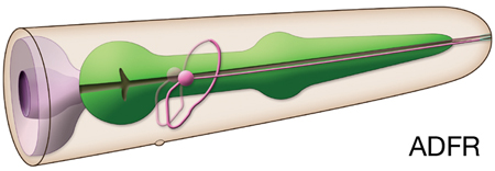
Neurotransmitter/ Neuropeptide:
- Acetylcholine
- Serotonin
- FLP-6; FMRFamide-like peptide
- INS-1; insulin-like peptide
- NLP-3; neuropeptide-like peptide
(Pereira et al., 2015; Loer, 2010; Li and Kim, 2008; Kodama et al., 2006; Nathoo et al., 2001; Sze et al., 2000; Duerr et al.,1999)
Innexin expression:
- INX-4 (in early larva)
- INX-19
(Altun et al., 2009; Chuang et al., 2007)
Receptor expression:
- MGL-3; metabotropic glutamate receptor family protein
- NPR-5; receptor for flp-3 and flp-18 encoded peptides
- OSM-9; TRPV (transient receptor potential channel, vanilloid subfamily; mammalian capsaicin receptor-like channel)
- OCR-2; TRPV (transient receptor potential channel, vanilloid subfamily; mammalian capsaicin receptor-like channel)
- SRB-6 (faint); G protein-coupled seven transmembrane receptor
- SRD-1 (in male); G protein-coupled seven transmembrane receptor
- TMC-1; putative cation channel, salt-sensing receptor
(Wormbase; Chatzigeorgiou et al., 2013; Altun, 2011; Cohen et al., 2009; Greer et al., 2008; Tobin et al., 2002; Colbert et al., 1997; Troemel et al.,1995)
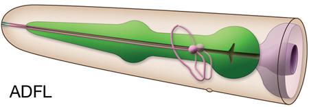
Function: ADFL/R are the only serotonergic sensory neurons in the hermaphrodite worm (in males, ray sensory neurons 1B, 3B and 9B are also serotonergic) and may couple environmental food signals with serotonin neurotransmission (Jafari et al., 2011)
- They contribute to a residual chemotactic response to cAMP, biotin, Cl-, and Na+ after ASE is killed (Bargmann and Horvitz, 1991). C. elegans shows chemoattraction to low NaCl. This is mediated by four pairs (ADF, ASE, ASG and ASI) of amphid sensory neurons, of which the ASE cells are most important. However, under hypoxic conditions, an additional, latent circuit involving ADF and ASG is activated for processing this chemosensory information (Pocock and Hobert, 2010.) C. elegans also shows avoidance of NaCl concentrations above 200 mM which is thought to be due to a general avoidance of high osmotic strength (mainly mediated by the ASH sensory neurons). However, this response to salt is plastic, involving a balance between attraction and avoidance, i.e., balances between ASE, ASI, ASH, ADF and perhaps ADL neuron (Hukema et al, 2006).
- Control entry into dauer stage; ADF, ASI and ASG inhibit entry into dauer stage while ASJ and ASK promote dauer entry (Kim et al., 2009; Ouellett et al., 2008; Schackwitz et al., 1996; Bargmann and Horvitz, 1991).
- Function as part of the oxygen-sensing network (which also includes URX, AQR, PQR, SDQ, ALN, PLN, ADL, and ASH) by promoting hyperoxia avoidance. ADF pair stimulates aerotaxis in the absence of food by producing serotonin (Chang et al., 2006).
- Modulate NMJ neurotransmission; serotonin functions as a neuromodulator by inhibiting and enhancing synaptic transmission of other neurotransmitters; e. g. studies have revealed both stimulatory and inhibitory serotonin inputs to the NMJs. Aldicarb, an inhibitor of acetylcholinesterase, causes paralysis in C. elegans due to accumulation of acetylcholine (ACh) at the locomotory NMJs, but exogenous serotonin inhibits paralysis induced by aldicarb. By contrast, exogenous serotonin does not reduce the paralysis induced by levamisole, a specific agonist of the nicotinic ACh receptor UNC-29 in the bodywall muscles. This suggests that serotonin signaling inhibits ACh release by the motor neurons. Studies showed that the inhibitory and stimulatory serotonin signals to NMJ neurotransmission arise from distinct serotonergic neurons; endogenous serotonin released from the ADF neurons stimulates ACh synaptic transmission at the NMJs, whereas serotonin from the NSM/RIH/AIM neurons inhibits the stimulatory serotonin inputs (Govorunova et al., 2010). |


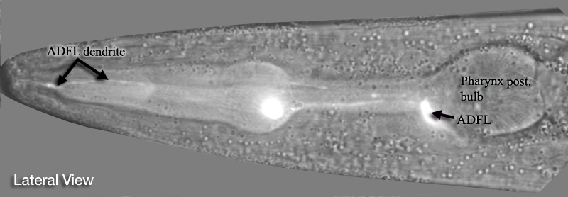
 Click pictures for higher resolution images
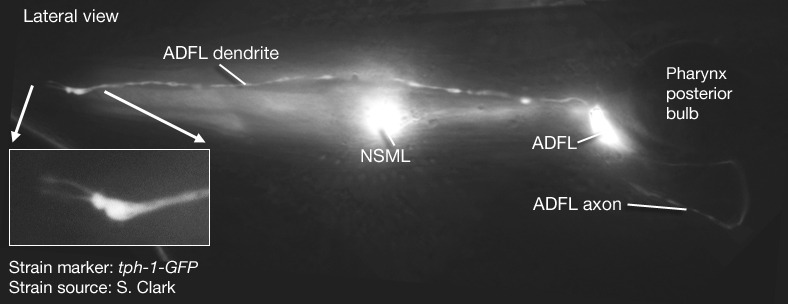