|
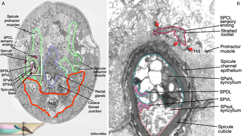
MaleSpicFIG 6: Ultrastructure of the spicule channel.
A. Low-power TEM showing the spicule channel and its relationship with the spicule muscle cells, transverse view. (Image source: N2Y [MRC] 60.)
B. Higher magnification TEM of white boxed region in A, transverse view. (Image source: N2Y [MRC] 984L.) (PAG) Pre-anal ganglion; (Hd) hemidesmosome. |