1 Spicule Muscles: the Retractors, Protractors and Anal Depressor
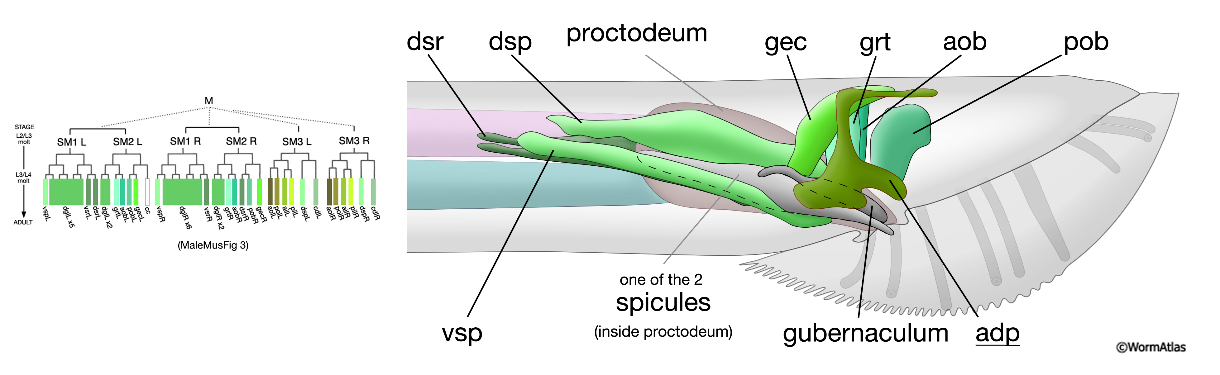
The male copulatory spicules are associated with three sets of muscles (MaleMusFIG 21 and 22): (1) the dorsal and ventral spicule retractor muscles, dsrL/R and vsrL/R; (2) the spicule protractor muscles, dspL/R and vspL/R; (3) the anal depressor, adp. The retractor and protractor muscles are male-specific and are generated post-embryonically by the M lineage. The anal depressor is a sexually dimorphic muscle, present in both sexes but specialized in the male to function as an auxiliary spicule muscle (Sulston et al., 1980; see Male Muscles - Overview).
MaleMusFIG 21: The spicule muscles. Right panel: Illustration of the adult male tail region featuring the spicules, left lateral view. Sexually dimorphic muscles are underlined. (adp) anal depressor, (aob/pob) anterior and posterior oblique muscles, (v/dsrL) ventral and dorsal spicule retractors, (v/dspL) ventral and dorsal spicule protractors; (gec) gubernacular erector. Left panel; Lineage tree of male muscles (as shown in MaleMusFIG 3)
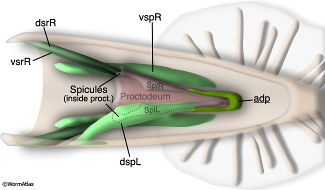
MaleMusFIG 22: The spicules. Illustration of the adult male tail region featuring the spicules, dorsal view. Sexually dimorphic muscles are underlined.(v/dsrL) Ventral and dorsal spicule retractors,(v/dspL) ventral and dorsal spicule protractors; (adp) anal depressor. (Adapted from Sulston et al., 1980.)
1.1 Spicule Retractor Muscles
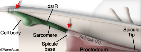
Two retractor muscles, one dorsal (dsrL/R) and one ventral (vsrL/R), attach to the base of each spicule. At their opposite ends the muscles attach to the sublateral body wall (MaleMusFIG 22, 23 and 24; Sulston et al., 1980). Each muscle contains a single sarcomere that occupies most of the cell. The myofilaments run A/P and so contraction of these muscles pulls the spicules inside the body.
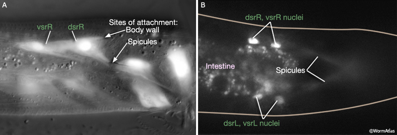
MaleMusFIG 23: Spicule retractor muscle attachment sites. A. Epifluorescent image from transgenic animals expressing the egl-15 ::GFP reporter gene (FGF receptor), lateral, right medial plane. (Strain source: C. Branda and M. Stern.) B. Epifluorescent image from transgenic animals expressing the hlh-8 ::GFP reporter gene (transcription factor), dorsal view. (Strain source: B.D. Harfe, M. Krause and A. Fire.)
MaleMusFIG 24: Spicule retractor muscle. Illustration featuring the male tail and spicule retractor muscles, dorsal view, right side.
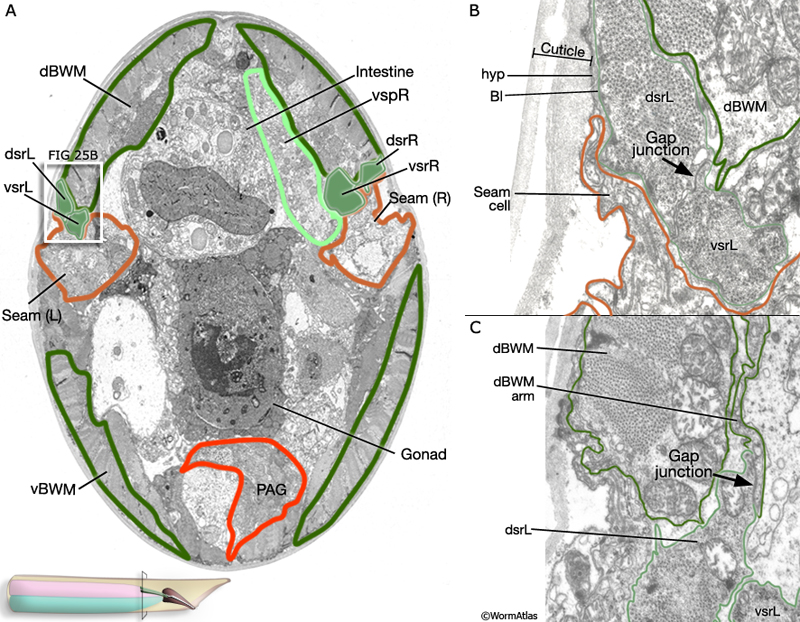
At their body wall end (MaleMusFIG 25A), dorsal and ventral retractors of a side are in direct contact for some distance along their length and are coupled by extensive gap junctions (MaleMusFIG 25B and 26). In addition, the dorsal retractors form gap junctions with dorsal body wall muscles (dBWM, MaleMusFIG 25C and 26; Sulston J.E., Albertson, D.G. and Thomson J. N., unpublished). The identity of neurons that innervate the spicule retractors is not yet known.
MaleMusFIG 25: Ultrastructure of the spicule retractors. A. Low-power TEM featuring the gap junctions coupling the dorsal and ventral retractor muscles, transverse section. (PAG) Pre-anal ganglion; (v/dBWM) ventral and dorsal body wall muscle; (v/dsrL/R) ventral and dorsal spicule retractors,(v/dspL) ventral and dorsal spicule protractors. (Image source: N2Y [MRC] 575-24.) B. TEM of boxed area in 25A showing close up of the gap junction between the dorsal and ventral spicule retractor. (hyp) Hypodermis; (Bl) basal lamina. (Image source: N2Y [MRC] 6824-24.) C. TEM showing close up of the gap junction between the dorsal spicule retractor and dorsal body wall muscle. (Image source: N2Y [MRC] 686-12.)
At the spicule end of the muscle (MaleMusFIG 26A), muscle::spicule attachment sites appear analogous to those observed between non-striated muscles and body wall cuticle. Hemidesmosomes (Hds) connect the muscle to the basal lamina, which in turn is connected to proctodeal epithelium. Proctodeal epithelium is attached to spicule cuticle (Sulston J.E., Albertson, D.G. and Thomson J. N., unpublished). These retractor muscle::spicule attachments do not appear to be as numerous or as tightly localized as those observed between protractor muscle and spicule (compare MaleMusFIG 26B with MaleMusFIG 29B).
MaleMusFIG 26: Spicule attachments to the retractors. A. Low-power TEM featuring the attachments between the retractor muscles and the spicules, transverse section. (PAG) Pre-anal ganglion; (v/dBWM) ventral and dorsal body wall muscle; (v/dsrL/R) ventral and dorsal spicule retractors,(v/dspL) ventral and dorsal spicule protractors; (Sph) sphincter. (Image source: N2Y [MRC] 596-24.) B. TEM of boxed area in 25A showing close up of the hemidesmosomes that connect the muscle to the basal lamina (Bl). (Image source: N2Y [MRC] 667-19.) C. Illustration depicting the gap junction connections between different muscle types. (Based on N2Y series, MRC.)
In addition to regulation of adult spicule behavior, the retractor muscles have a developmental role and are required for proper anterodorsal elongation of the proctodeum during development. In males that lack retractor muscles (or M) the proctodeum fails to elongate and its internal structures, the spicules and gubernaculum, are compressed (Sulston et al., 1980). During wild type larval development cells on the dorsal surface of the proctodeum insert "fingers" into the nearby retractor muscles. During male tail morphogenesis (late L4) anterior movement of the muscles (carried by the general forward movement of the body wall) draws out the proctodeum, the spicules and gubernaculum (Sulston et al., 1980).
1.2 Spicule Protractor Muscles
Two protractor muscles (one dorsal dspL/R; one ventral, vspL/R) are attached to the base of each spicule (MaleMusFIG 27; Sulston et al., 1980). These muscles, particularly the dorsal protractors, have large cell bodies. Each muscle contains a single sarcomere with myofilaments that run in the A/P orientation. The sarcomere is concentrated in the region of the muscle that overlaps with the spicules (MaleMusFIG 28). At their anterior edges, the dspL/R are attached to the dorsal sublateral body wall and at their posterior edge to the anal depressor (adp). The vspL/R are attached to the ventral sublateral body wall (MaleMusFIG 22). Contraction of the protractors therefore has the opposite affect to retractor contraction and causes the spicules to extrude from the body. |
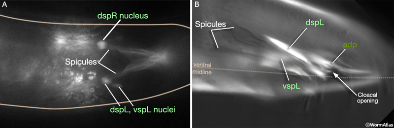
MaleMusFIG 27: Spicule protractor muscle attachment sites. A. Epifluorescent image from transgenic animals expressing the hlh-8 ::GFP reporter gene (transcription factor), sub-dorsal view. (Strain source: B.D. Harfe, M. Krause and A. Fire.) B. Epifluorescent image from transgenic animals expressing the unc-27::GFP reporter gene (Troponinl, thin filaments), left, lateral oblique view. (Strain source: L. Jia and S.W. Emmons.)
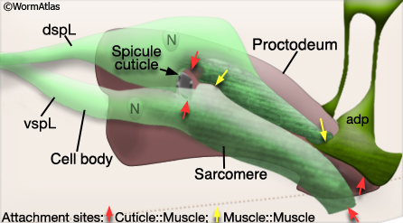
MaleMusFIG 28: Spicule protractor muscle. Illustration featuring the sarcomeres of the spicule protractor muscles, left lateral view.
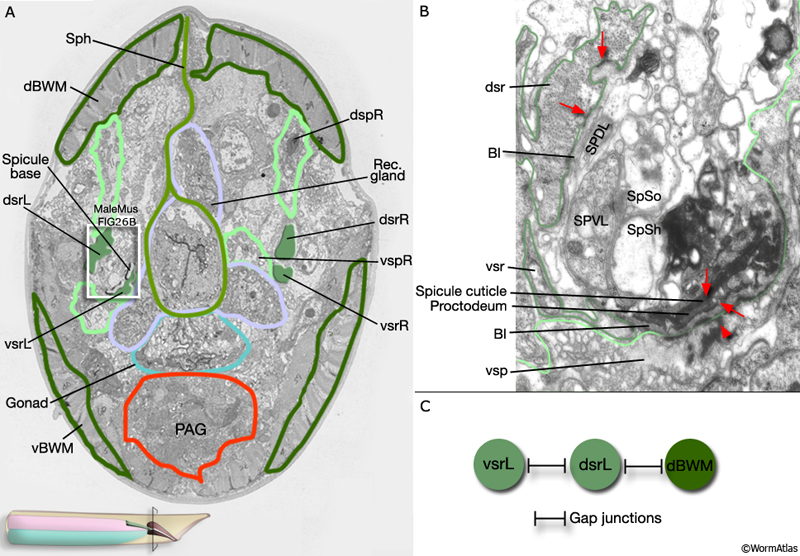
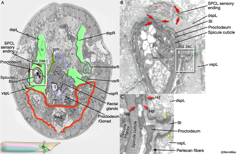
The protractor muscles are connected to the spicules via a series of hemidesmosomes (Hds) that interconnect muscle, basal lamina, proctodeum and spicule cuticle (MaleMusFIG 29A&B; red arrows). Attachment plaques link spicule muscles to each other: dorsal protractors to ventral protractors and dorsal protractors to anal depressor (MaleMusFIG 29C; yellow arrows).
MaleMusFIG 29: Connection of the protractor muscles to the spicules. A. Low-power TEM featuring the location of the spicules within the spicule protractor muscle, transverse section. (PAG) Pre-anal ganglion; (v/dsrL/R) ventral and dorsal spicule retractors, (v/dspL) ventral and dorsal spicule protractors. (Image source: N2Y [MRC] 597-5.) B. TEM of boxed area in 29A showing close up of the hemidesmosomes (red arrows) that interconnect muscle, basal lamina, proctodeum and spicule. (Bl) basal lamina. (Image source: N2Y [MRC] 517-4.) C. TEM of boxed area in 29B showing close up of the attachment plaques (yellow arrows) that link spicule muscles to each other, dorsal protractors to ventral protractors and dorsal protractors to the anal depressor. (Image source: N2Y [MRC] 517-4.)
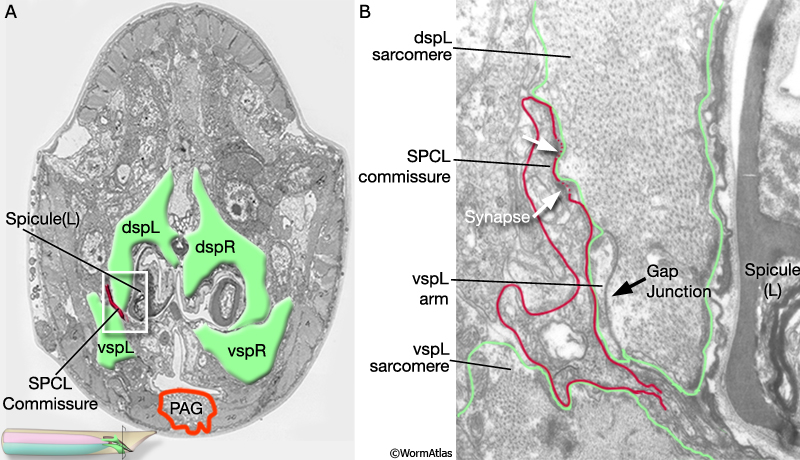
The protractor muscles are directly innervated by neurons SPCL/R (MaleMusFIG 30 and 31; Sulston et al., 1980). The SPCL/R neurons have both sensory (proprioceptive/mechanosensory) and motor neuron function. Their sensory endings are directly attached to the dorsal protractor muscles via hemidesmosomes (MaleMusFIG 29B and 30; Male Wiring Project). SPCL/R form numerous neuromuscular junctions (NMJs) with both dorsal and ventral muscles and a few with the anal depressor (Male Wiring Project). Interestingly, these synapses are not on muscle arms but on the main body of the muscle that contains the sarcomere (MaleMusFIG 31). Muscle activity is also regulated by the post-cloacal sensilla (PCS) neurons although this may be indirect as PCS neurons do not appear to synapse with spicule muscles (Male Wiring Project). SPCL/R and PCS neurons control different aspects of spicule behavior: PCS neurons, detecting the presence of the vulva, induce periodic contraction of the muscles which causes the spicules to prod the hermaphrodite surface for the vulval opening; SPCL/R stimulate prolonged contraction which causes the spicules to extrude fully and insert into the vulva once the opening is located. Both the PCS neurons and SPCL/R regulate muscle activity through release of acetylcholine. The gonad also influences the behavior of these muscles as its presence is required for maintaining prolonged muscle contraction so that spicules remain fully inserted during sperm transfer (Garcia et al., 2001; see MOVIE of spicule behavior by L. Rene Garcia)
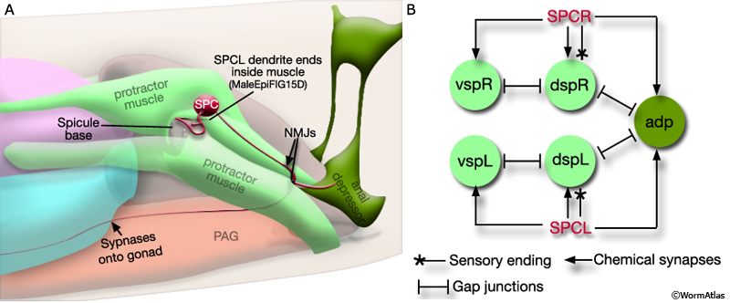
MaleMusFIG 30: Innervation of the protractor muscles. A. Illustration showing the innervation of the protractors by the SPC neurons, lateral view. (PAG) pre-anal ganglion; (NMJs) neuromuscular junctions. B. Diagram of the electrical connections of the neurons and muscles of the spicules. (Based on N2Y series, MRC.)
MaleMusFIG 31: Innervation of the protractor muscles. A. Low power electron micrograph featuring the coupling of the protractors and anal depressor, transverse view. (Image source: N2Y [MRC] 45.) B. Detailed view of boxed region in A showing synapses and gap junction. (PAG) Pre-anal ganglion; (v/dspL/R) ventral and dorsal spicule protractors.
The protractors and anal depressor are not only physically connected to each other and regulated by common neurons, they are also coupled electrically. The dspL/R and vspL/R muscles of each side are connected by gap junctions (MaleMusFIG 30B and 31A); dspL and dspR, in turn, are electrically coupled to the anal depressor (MaleMusFIG 30B and 32; Sulston J.E., Albertson, D.G. and Thomson J. N., unpublished). Electrical coupling in this way likely facilitates coordinated contraction of the spicule muscle (MaleMusMOVIE 2).
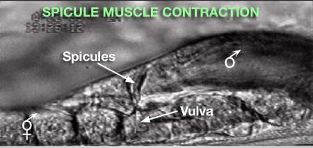
MaleMusMOVIE 2: Spicule muscle contraction. Periodic vs Prolonged Spicule Muscle Contraction (Image source: L. Rene Garcia.) Click on image to play movie.
1.3 Anal Depressor Muscle
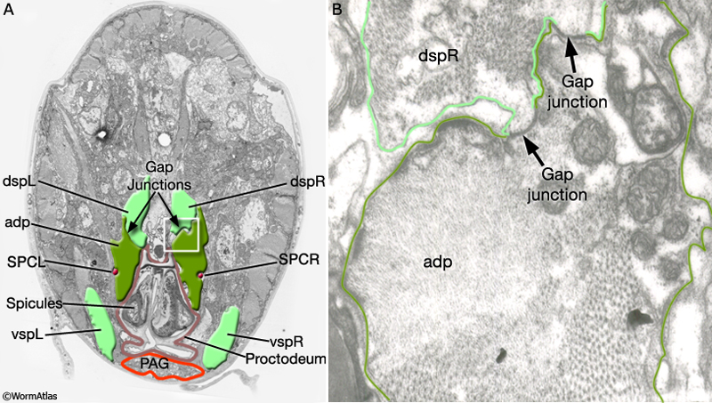
As described in Male Muscles - Overview, the anal depressor muscle (adp) is a sexually dimorphic muscle. In males, instead of participating in defecation, it functions as an auxiliary spicule protractor muscle (MaleMusFIG 30A and 32; Sulston et al., 1980; Garcia et al., 2001). The muscle contains a single sarcomere with myofilaments oriented the same direction as the dspL/R i.e. A/P. Along its ventral edges the adp is attached to the proctodeum/gubernaculum and to the ventral body wall. Dorsally it retains some connections sublaterally to the dorsal roof (MaleMusFIG 30A). Where adp and the dspL/R overlap there are extensive regions of gap junctions (MaleMusFIG 32; Sulston J.E., Albertson, D.G. and Thomson J. N., unpublished).
MaleMusFIG 32: Anal depressor muscle. A. Low power electron micrograph featuring anal depressor and gap junctions to the protractor muscles, transverse view. (Image source: N2Y [MRC] 37.) B. Detailed view of boxed region in A showing gap junctions. (PAG) Pre-anal ganglion; (v/dspL/R) ventral and dorsal spicule protractors; (adp) anal depressor.
2 Gubernacular Muscles
The gubernaculum is a thick sclerotic strip of cuticle that lines the roof of the cloaca and is thought to guide the movement of spicules ventrally through the anus (described in detail in The Proctodeum; Sulston et al., 1980). Two bilateral pairs of sex-specific muscles are attached to the gubernaculum: (1) the gubernacular erectors (gecL/R) and (2) the gubernacular retractors (grtL/R) (MaleMusFIG 33 and 34; Sulston et al., 1980). Both muscle types contain a single sarcomere with myofilaments oriented dorsoventrally.
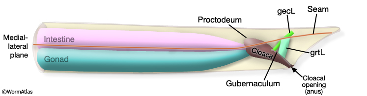
MaleMusFIG 33: Gubernacular muscles. Illustration of the adult male tail region featuring the gubernaculum and gubernacular muscles, left lateral view. (gecL/R) Gubernacular erectors; (grtL/R) gubernacular retractors.
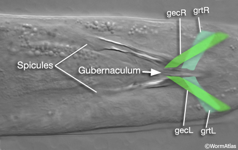
MaleMusFIG 34: Gubernacular muscles. Nomarski DIC showing the location of the gecL/R and grtL/R muscles within the gubernaculum, dorsal view. (gecL/R) Gubernacular erectors; (grtL/R) gubernacular retractors.
2.1 Gubernacular Erector Muscles
The gubernacular erectors (gecR/L) attach at their ventral edges to the gubernaculum and cloacal roof (MaleMusFIG 35). At their dorsal edge these muscles attach to the dorsal sublateral body wall.
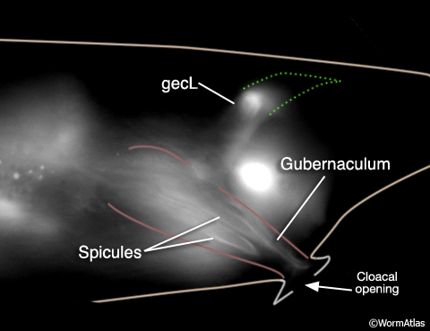
MaleMusFIG 35: Gubernacular erector muscles. Epifluorescent image from transgenic animal expressing the egl-15 reporter gene (FGF receptor), lateral right medial plane. The gubernacular erectors attach to the gubernaculum and cloacal roof by their ventral edges and to the dorsal sublateral wall by their dorsal edge. (Strain source: C. Branda and M. Stern.)
The gubernacular erectors (gecL/R) are innervated on their muscle arms by 2 of the PCS (post-cloacal sensilla) neurons, PCAL/R and the left PCB neuron, PCBL (MaleMusFIG 36; Male Wiring Project). After the PCS and/or hook detect the approximate location of the vulva, PCS neurons trigger spicule prodding behavior which is used to pinpoint the vulval opening (Liu and Sternberg, 1995; Garcia et al., 2001). At this time the gubernaculum twitches possibly due to PCS neuron-stimulated contraction of the gec muscles. During ejaculation, gubernaculum muscle contraction pulls the male proctodeum/protracted spicules posteriorly to allow the opening of the vas deferens access to the cloacal opening. Laser-ablation of the gubernaculum muscles causes the protracted spicules to block the vas deferens, thus inhibiting sperm release (Liu et al., 2011).
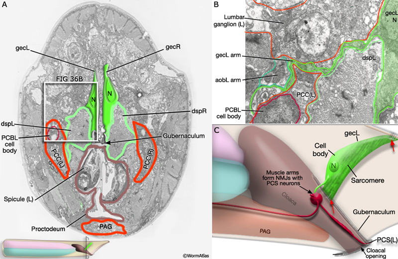
MaleMusFIG 36: Innervation of the gubernacular erectors. A. Low-power TEM showing the innervation of the gubernacular erectors by the post cloacal sensilla neurons (PCB and PCC), transverse section. (PAG) Pre-anal ganglion; (gecL/R) gubernacular erectors; (grtL/R) gubernacular retractors; (dspL/R) dorsal spicule protractors. (Image source: N2Y [MRC] 597-5.) B. TEM of boxed area in 36A showing close up of the lumbar ganglion and gubernacular muscles, basal lamina, proctodeum and spicule. (Bl) basal lamina. (Image source: N2Y [MRC] 559-6 [N2YLP41].) C. Illustration of the gubernacular and surrounding region, left lateral view.
2.2 Gubernacular Retractor Muscles
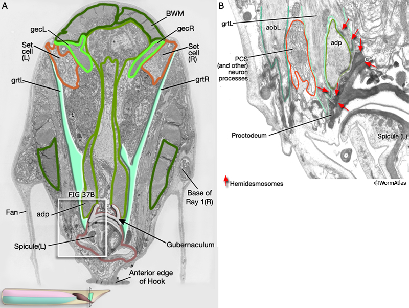
The gubernacular retractor (grtL/R) muscles attach at their ventral edges to the gubernaculum and overlying proctodeal epithelium. At their dorsal edges the muscles attach to the sublateral dorsal body wall, immediately below the lateral seam (called the set cell in this region in the male) (MaleMusFIG 37). The neurons that innervate the gubernacular retractors have not yet been identified.
MaleMusFIG 37: Gubernacular retractor muscles. A. Low-power TEM featuring the location gubernacular retractor muscles and their ventral attachment to the gubernacular and proctodeal epithelium. (BWM) Body wall muscle; (gecL/R) gubernacular erectors; (grtL/R) gubernacular retractors; (adp) anal depressor. (Image source: N2Y [MRC] 597-5.) B. TEM of boxed area in 37A showing close up of the hemidesmosomes (red arrows) in this region. (Image source: N2Y [MRC] 581-1.)
3 Oblique Muscles
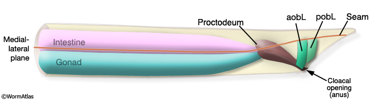
Two bilateral pairs of sex-specific (M-derived) oblique muscles are located in the anal region of the adult male tail; an anterior pair (aobL/R) and a posterior pair (pobL/R) (MaleMusFIG 38, 39 and 40; Sulston et al., 1980). These muscles contain a single sarcomere oriented dorsoventrally. At their dorsal edge, the muscles attach to the body wall, immediately below the set cell; along their ventral edge the muscles attach ventral body wall.
MaleMusFIG 38: Oblique muscles of the male tail. Illustration of the adult male tail region featuring the proctodeum and oblique muscles, left lateral view. (aobL/R) anterior oblique muscle; (pobL/R) posterior oblique muscle.
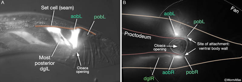
MaleMusFIG 39: Sex-specific oblique muscles. A. Epifluorescent image from transgenic animal expressing the egl-15 reporter gene (FGF receptor), left lateral oblique view. B. Epifluorescent image showing the attachment sites of the oblique muscles to the ventral body wall. (aobL/R) Anterior oblique muscle; (pobL/R) posterior oblique muscle.
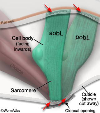
MaleMusFIG 40: Oblique muscles with cell bodies. Illustration featuring the oblique muscles and their attachment sites to the body wall along their dorsal edge and to the ventral body wall below the Set cell along their ventral edge, left lateral view. (aobL/R) Anterior oblique muscle; (pobL/R) posterior oblique muscle.
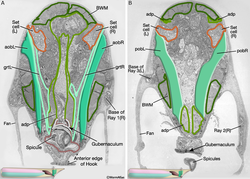
MaleMusFIG 41: Ultrastructure of oblique muscles. A. Low power TEM, transverse view of male tail region featuring the anterior oblique muscles. (Image source: N2Y [MRC] 598-9.) B. Low power TEM, transverse view of male tail region, further posterior to region shown in A and featuring the posterior oblique muscles (Image source: N2Y [MRC] 598-14.) (aobL/R) Anterior oblique muscle; (pobL/R) posterior oblique muscle; (BWM) body wall muscle; (gecL/R) gubernacular erectors; (grtL/R) gubernacular retractors; (adp) anal depressor.
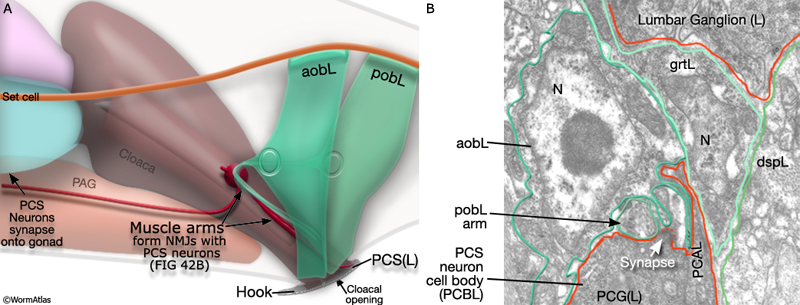
Anterior and posterior oblique muscles on each side receive synaptic inputs from all three post-cloacal sensillum (PCS) neurons, namely PCAL/R, PCBL/R and PCCL/R (Male Wiring Project). Synapses are located on muscle arms that project from the muscle cell body and run with PCS neuron processes as they descend towards the left or right sensillum (MaleMusFIG 42A&B; Sulston J.E., Albertson, D.G. and Thomson J. N., unpublished). As mentioned above, the PCS neurons trigger spicule muscle contraction in response to a vulval cue (Liu and Sternberg, 1995; Garcia et al., 2001). PCS neuron-stimulated contraction of the oblique muscles changes the posture of the tail, and thus, help the male press the posterior region of his tail over the vulva during prodding attempts.
MaleMusFIG 42: Synaptic input to the oblique muscles. A. Illustration showing the locations of the NMJs between the PCS neurons and the oblique muscles, left lateral view. B. TEM of NMJ region featured in A showing a synapse between PCG and oblique muscle. (VNC) Ventral nerve cord; (vBWM) ventral body wall muscle. (Image source: N2Y [MRC] 595-19.) (aobL/R) Anterior oblique muscle; (pobL/R) posterior oblique muscle; (grtL) gubernacular retractor; (dspL) dorsal spicule protractor; (N) nucleus.
4 Sphincter Muscles
As described in Male Muscles - Overview, the sphincter (sph), like the anal depressor, is a sexually dimorphic muscle, common to both sexes but modified during male larval development (MaleMusFIG 43).
MaleMusFIG 43: Sphincter muscles. Illustration of the adult male tail region featuring the sphincter muscle (sph), left lateral view. Sexually dimorphic muscles are underlined.
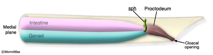
In the adult male the alimentary tract and reproductive tract are joined to a common chamber, the cloaca, which opens to the environment at the anus (described in detail in The Proctodeum). In contrast to the hermaphrodite anus, the male cloaca is constitutively open presumably to permit free movement of the spicules (housed within) and sperm during mating behavior. The principal seal for the male alimentary tract is instead at the sphincter (MaleMusFIG 44).
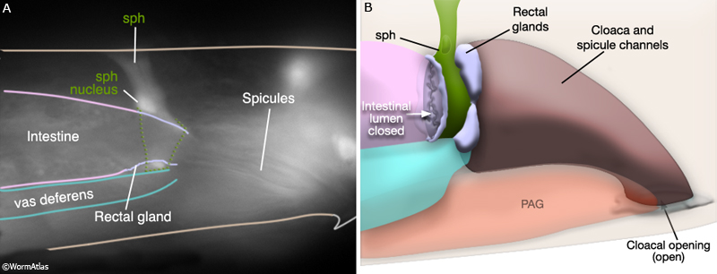
MaleMusFIG 44: Positioning of the sphincter. A. Epifluorescent image from transgenic animal expressing the egl-15 reporter gene (FGF receptor), lateral right medial plane. The sphincter muscle (sph) and nucleus are proximate to the spicules and rectal gland. (Strain source: C. Branda and M. Stern.) B. Diagram showing the position of the sphincter muscle in the male tail. (PAG) Pre-anal ganglion.
The male sphincter is tonically contracted and must be relaxed (through inhibitory GABA transmission) during defecation. This response to GABA is opposite to that observed in larval males and in adult hermaphrodites and may reflect a change in receptor expression in the muscle (Reiner and Thomas, 1995). The male sphincter also differs morphologically from larval male and hermaphrodite sphincters. It is enlarged (hypertrophied) but still contains a single sarcomere. It has a dorsal process that attaches to the dorsal roof and that contains myofilaments (MaleMusFIG 45). Thus hyper-contraction of the sphincter during ejaculation not only clamps off the intestine but also draw it away from the vas deferens, possibly allowing sperm to pass more freely (Sulston et al., 1980).
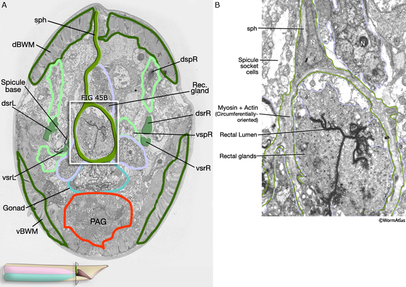
MaleMusFIG 45: Ultrastructure of the sphincter muscle. A. Low-power TEM featuring the sphincter muscle. (PAG) Pre-anal ganglion; (v/dsrL/R) ventral and dorsal spicule retractors, (v/dspL) ventral and dorsal spicule protractors; (d/vBWM) dorsal and ventral body wall muscle. (Image source: N2Y [MRC] 596-24.) B. TEM of boxed area in 45A showing close up of the myosin and actin filaments within the sphincter muscle. (N) Nucleus. (Image source: N2Y [MRC] 942-L.)
5 List of Male Specific Muscle Cells (See M lineage and MaleMusFIG 3)
Diagonal (dgl) muscles of the adult
SM1L.aap
SM1L.apa
SM1L.app
SM1L.paa
SM1L.pap
SM2L.aaa
SM2L.aap
SM1R.aap
SM1R.apa
SM1R.app
SM1R.paa
SM1R.pap
SM1R.ppa
SM2R.aaa
SM2R.aap
Ventral longitudinal muscles of the adult
1. Outer longitudinal
SM3R.aap (polR, posterior outer longitudinal, right)
SM3R.aaa (aolR, anterior outer longitudinal, right)
SM3L.aap (polL, posterior outer longitudinal, left )
SM3L.aaa (aolL, anterior outer longitudinal, left)
2. Inner longitudinal
SM3R.app (pilR, posterior inner longitudinal, right)
SM3R.apa (ailR, anterior inner longitudinal, right)
SM3L.app (pilL, posterior inner longitudinal, left)
SM3L.apa (ailL, anterior inner longitudinal, left)
3. Caudal longitudinal
SM3R.pp (cdlR, right)
SM3L.pp (cdlL, left)
Spicule muscles of the adult
1. Spicule protractors
SM3R.pa (dspR, dorsal right)
SM1R.aaa (vspR, ventral right)
SM3L.pa (dspL, dorsal left)
SM1L.aaa (vspL, ventral left)
2. Spicule retractors
SM2R.paa (dsrR, dorsal right)
SM1R.ppp (vsrR, ventral right)
SM1L.ppp (dsrL, dorsal left)
SM1L.ppa (vsrL, ventral left)
Gubernacular muscles of the adult
1. Gubernacular erectors
SM2R.pp (gecR, right)
SM2L.pap (gecL, left)
2. Gubernacular retractors
SM2R.apa (grtR, right)
SM2L.apa (grtL, left)
Oblique muscles of the adult SM2R.pap (pobR, posterior right)
SM2R.app (aobR, anterior right)
SM2L.paa (pobL, posterior left)
SM2L.app (aobL, anterior left)
Anal sphincter muscle (See Alimentary System of the Male)
mu sph
Anal depressor muscle
mu anal
6 References
Branda, C.S. and Stern, M.J. 2000. Mechanisms controlling sex myoblast migration in Caenorhabditis elegans hermaphrodites. Dev. Biol. 226: 137-51. Article
Garcia, L.R., Mehta, P. and Sternberg, P.W. 2001. Regulation of distinct muscle behaviors controls the C. elegans male's copulatory spicules during mating. Cell 107: 777-788. Article
Liu, K.S. and Sternberg, P.W. 1995. Sensory regulation of male mating behavior in Caenorhabditis elegans. Neuron 14: 79-89. Article
Liu, Y., LeBeouf, B.,Guo, X., Correa, P.A., Gualberto, D.G., Lints, R. and Garcia, L.R. 2011. A cholinergic-regulated circuit coordinates the maintenance and bi-stable states of a sensory-motor behavior during Caenorhabditis elegans male copulation. PLoS Genetics 7;e1001326. Article
Reiner, D.J. and Thomas, J.H. 1995. Reversal of a muscle response to GABA during C. elegans male development. J. Neurosci. 15: 6094-6102. Article
Sulston, J.E., Albertson, D.G. and Thomson, J.N. 1980. The Caenorhabditis elegans male: Postembryonic development of nongonadal structures. Dev Biol. 78: 542-576. Article
|

 Click pictures for new window with figure and legend