|
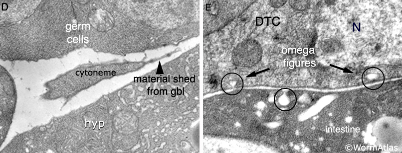
SomaticFIG 2D&E: Transmission electron micrograph images of the DTC.
D. Longitudinal section, shows fragments of the gbl shed from the trailing arms of the DTC into the pseudocoelom. (Hyp) Hypodermis;
(GBL) Gonadal basal lamina. (Image source: Hall archive.)
E. Longitudinal section, from an area of the plasma membrane that sometimes displays "omega"figures where it faces the gbl, indicative of active endocytosis or endocytosis. (N) nucleus. (Image source: Hall archive.)
See also SomaticFIG 2A.
Click on picture for full resolution image.
|