|
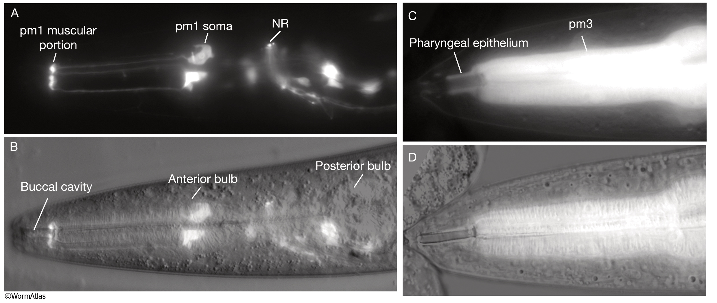
PhaFIG 4A-D: The anterior digestive track is formed by the alignment of a series of cells.
A&B. Sarcomeres of pm1 lie near the posterior edge of the buccal cavity. A. Epifluorescent image of pm1 in an animal expressing a GFP-tagged transgene. (NR) Nerve ring. (Strain source: Z-W. Wang and B. Chen.) Magnification 400x. B. Same animal in merged DIC and epifluorescent images.
C&D. Anterior to pm1, pharyngeal epithelium surrounds the posterior buccal cavity. C. Epifluorescent image of the pharyngeal epithelium and pm3 in an animal expressing a GFP-tagged transgene. (Strain source: Z-W. Wang and B. Chen.) Magnification, 600x. D. Same animal in merged DIC and epifluorescent images.
See also PhaFIG 4E-I.
Click on picture for full resolution image.
|