|
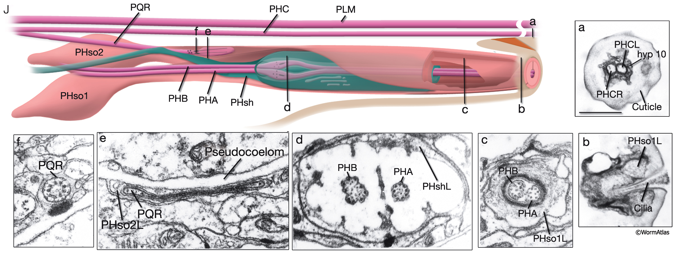
NeuroFIG 41J: Structure of the phasmid sensilla.
J. Left phasmid sensillum in the adult hermaphrodite. Also seen are processes of nonsensillar PHC and PLM neurons. (Top left panel) Schematic illustration indicating the section levels for insets a–f (all transverse-section TEMs, viewed from the caudal). a. Section through extreme tail tip with PHCL/R processes terminating embedded in hyp 10. Bar, 0.5 μm. b. Left phasmid opening with PHA and PHB cilia exposed to the outside. c. PHA and PHB cilia within the socket channel. d. Transition zones of PHA and PHB cilia with nine doublet MTs on the outside and singlet MTs on the inside. At this level, the channel is formed by the sheath cell. e. PQR ciliary ending within PHso2L. f. Transition zone of the PQR cilium. (Image source: [Hall] B136.)
See also NeuroFIG 4A-I
Click on picture for full resolution image.
|