|
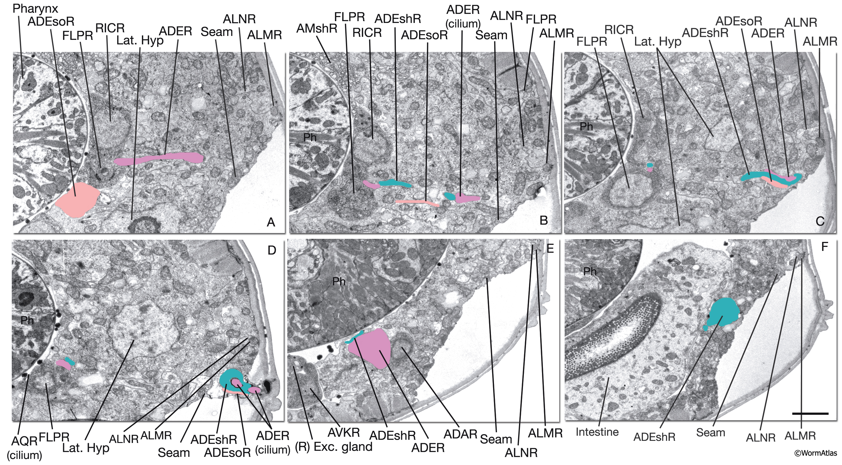
NeuroFIG 38: Cells of the right anterior deirid sensillum.
Low-power TEM images of the right anterior deirid, transverse views. The sensillar components are colored on the TEM images. All components lie medially to the lateral hypodermis. Sections are ordered from anterior to posterior throughout. (Ph) Pharynx. Bar, 1 μm.
A. Section through the cell body of the ADE socket cell. Also seen is the commissural (ventrally directed) process of ADER. (Image source: N2U [MRC] 217-19.)
B. Section through the processes of the socket and sheath cells and the ADER cilium as they travel together laterally. (Image source: N2U [MRC] 217-24.)
C. Section through the sensillar processes as they reach the outermost body wall. (Image source: N2U [MRC] 22017.)
D. Section through the ADER sensillum where sheath surrounds the cilium. (Image source: N2U [MRC] 221-6.)
E. Section through the cell body of the ADER neuron located on the lateral side of the terminal bulb of the pharynx. (Image source: N2U [MRC] 234-10.)
F. Section through the cell body of the ADEshR, which is located more posteriorly near the pharyngeal–intestinal valve. (Image source: N2U [MRC] 250-10.)
Click on picture for full resolution image.
|