|
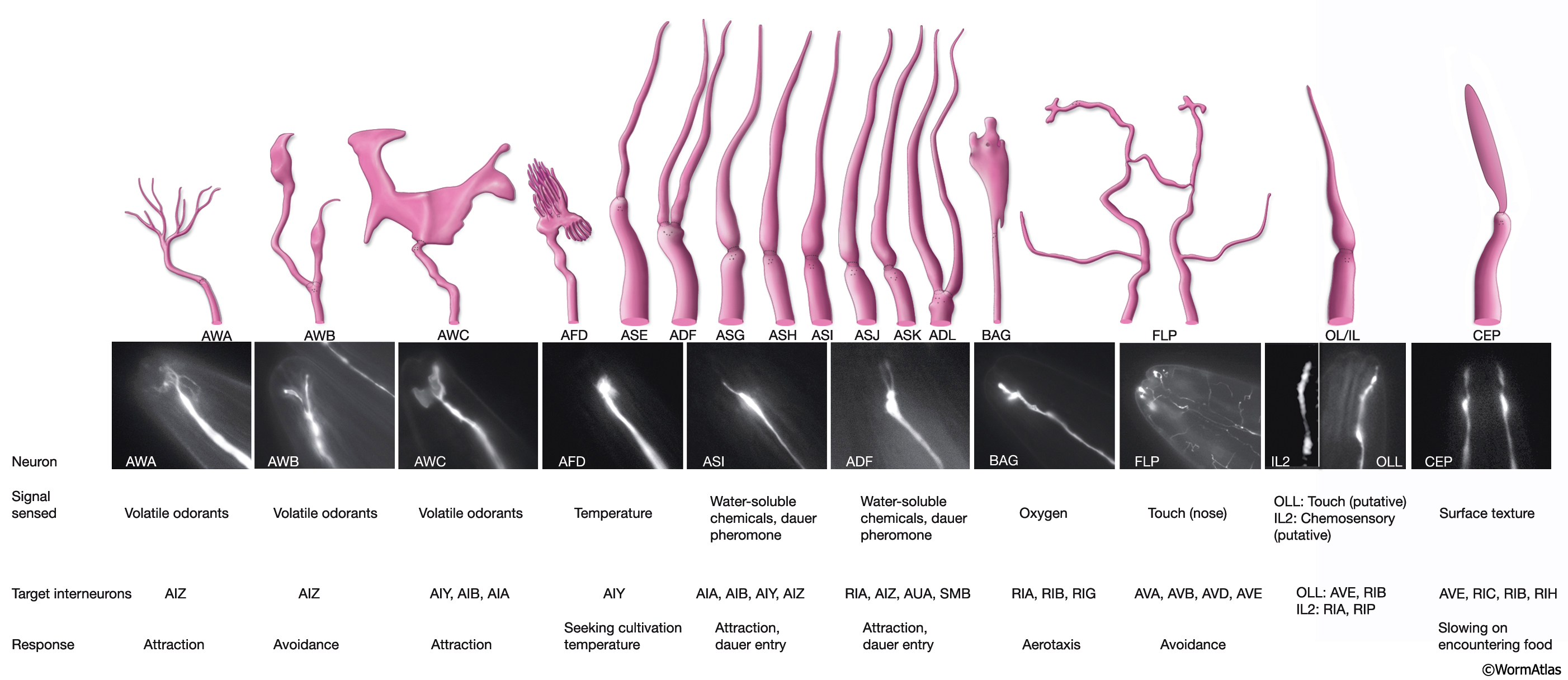
NeuroFIG 25: Cilia morphology of the sensory neurons that terminate in the lips.
(Based on Ward, 1973.) (Top panel) Illustrations of cilia. (Middle panel) Epifluorescent images of cilia (OLQ and IL1 are not shown). Although all chemosensory amphid cilia are similar in shape with one (ASE, ASG, ASH, ASI, ASJ, ASK) or two (ADF, ADL) slim, finger-like cylinders, each of the amphid wing cilia (AWA, AWB, AWC) has a unique shape (Ward et al., 1975; Ware et al., 1975). The AWC cilium spreads vertically into a large, wing-like sheet that fills up most of the lateral side of the sheath cell. AWA and AWB cilia are smaller and comparable in size to the chemosensory cilia. The distal segment of the AWA cilium arborizes into several small fingers. The AWB dendrite splits into a pair of cilia that becomes flattened and irregular at the distal regions. The AFD dendrite ends in a brush-like structure with many villi surrounding one cilium. BAG neurons terminate in an enlarged process distally to the cilium (Ware et al., 1975). The FLP cilium (the ending of a single cell is shown) is located at the distal end of its lateral process. Several branches extend dorsally and ventrally from the more proximal part of the dendrite. Each IL and OL dendrite terminates in a finger-like cilium. The CEP cilium widens as it ends in the cuticle. (Bottom panel) Table of the sensory cues detected by these neurons, the interneurons with which they synapse, and the behavioral response they elicit. Strain markers (strain source): AWA: odr-10::GFP (P. Sengupta), AWB: str-1::GFP (C. Bargmann/CGC), AWC: str-2::GFP (C. Bargmann/CGC), AFD: gcy8::GFP (P. Sengupta), ASI: sra-6::GFP (P. Sengupta), ADF: tph-1::GFP (S. Clark), BAG: gcy-33::GFP (L. Avery/CGC), FLP: mec-3::GFP (M. Chalfie), OLL: ser-2prom3::GFP (O. Hobert), and CEP: dat-1::GFP (S. Clark). Magnification, 600x. IL2: DiO labeling. (Image source: L. Ryder.)
Click on picture for full resolution image.
|