|
NeuroFIG 19: Nerve tracts and commissures of the head.
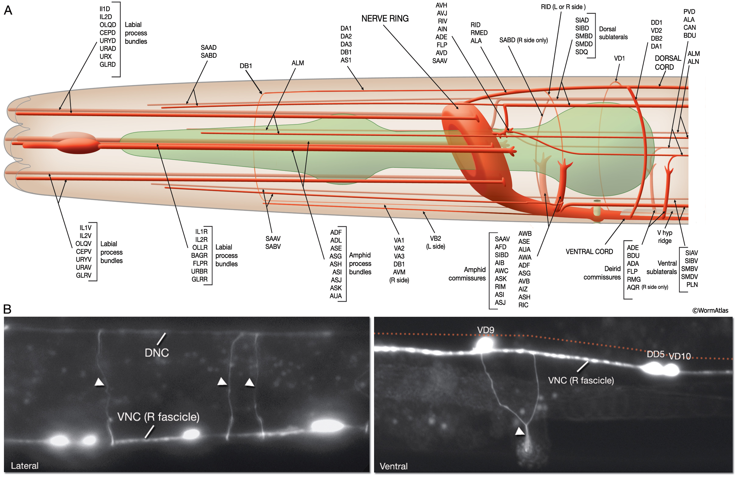
A. Longitudinal and circumferential fiber tracts in the head, left lateral view. Six labial process bundles are composed of the dendrites of the cephalic, inner, and outer labial neurons. Two amphid process bundles consist of amphid neuron dendrites and run on the lateral sides of the head. Other neuron processes, including BAG, FLP, URB, GLR, URY, URA, and URX, which have ciliated and non-ciliated endings, also travel within the labial nerve bundles and terminate in the anterior head or lips. The labial neuron axons run posteriorly within the labial nerves, pass along the outside of the ring, and then turn anteriorly and enter the NR to make synapses. Four minor tracts run subdorsally or subventrally and contain processes from SAA and SAB neurons. Anterior to the nerve ring, dorsal and ventral processes from the VNC motor neurons travel anteriorly in close association with dorsal and ventral hypodermal ridges. Posterior to the nerve ring, circumferential tracts follow lateral routes on the right or left sides between the dorsal and ventral levels (see NeuroFIG 13 and NeuroFIG 14). (Modified, with permission of The Royal Society of London, from White et al., 1986.)
B. Epifluorescent images from transgenic animals expressing the GABAergic neuron-specific unc-47::GFP reporter gene. The commissures (arrowheads) of the VNC motor neurons mostly travel on the right side of the body as single processes or as thin bundles of a few processes, lateral (top panel) and ventral (bottom panel) views. Anterior is to the left. Note that the left fascicle of the VNC (red dotted line) is not visible in these images, because the neurons that comprise (L) VNC do not express the GFP marker of this strain. The D-type motor neuron cell bodies are visible along the VNC, close to the right fascicle. Magnification, 600x. (Strain source: K. Schuske and E. Jorgensen.)
Click on picture for full resolution image.
|