|
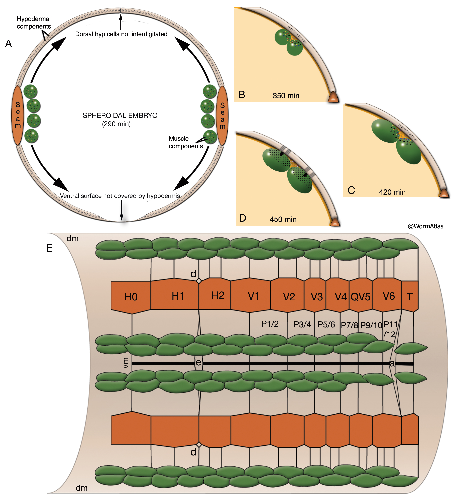
MusFIG 14A-E: Development of somatic muscle.
A. A 290-minute embryo after first cell cleavage. Myoblasts arise after the end of gastrulation and are localized adjacent to seam cells. They have started to accumulate structural components such as myosin, actin, vinculin, and integrin (small speckles inside myoblasts). Early myoblasts are almost spherical. The dorsal and ventral hypodermis contain some hemidesmosome components (dotted areas within the beige circle). Dorsal hypodermal cells have not yet intercalated.
B. A 350-minute embryo. Starting at the anterior end, muscle cells have migrated to form the ventral and dorsal muscle quadrants between 300 and 350 minutes and have reached their final positions. All muscle cells have gone through their final divisions. Muscle cells are still round but have become polarized; myofibrillar components (colored dots within muscle cells) are clustered where the two muscle cells appose each other and the hypodermis.
C. A 420-minute embryo. Muscle cells have flattened basally against the hypodermis and laterally against their muscle neighbors. Myofibrillar components, basal lamina, and hypodermal hemidesmosome components have aligned.
D. A 450-minute embryo, midembryogenesis. Muscle–muscle and muscle– hypodermis junctions and sarcomeres organize to provide a physical linkage between cells. All types of attachments (DB, M lines, attachment plaques) appear morphologically similar to electron-dense plaques when they are first formed (Coutu Hresko et al., 1994; Moerman and Williams, 2006). In larval stages, DBs and M lines acquire their finger-like shapes by extending deeper into the cytoplasm.
E. Schematic drawing of localization of embryonic somatic muscle cells (green) with respect to the hypodermis (beige) and seam blast cells (dark orange) in a filleted embryo. At the stage shown, the dorsal hypodermal cells have completed their intercalation and are organized in a single dorsal row. (a) Anus; (d) anterior deirid; (dm) dorsal midline; (e) excretory pore; (vm) ventral midline. Only the right-side seam blast cells and P cells are labeled. Hypodermal cells are unlabeled and the anterior hypodermis is omitted. (Based on Hedgecock et al., 1987.)
See also MusFIG 14F-K.
Click on picture for full resolution image.
|