|
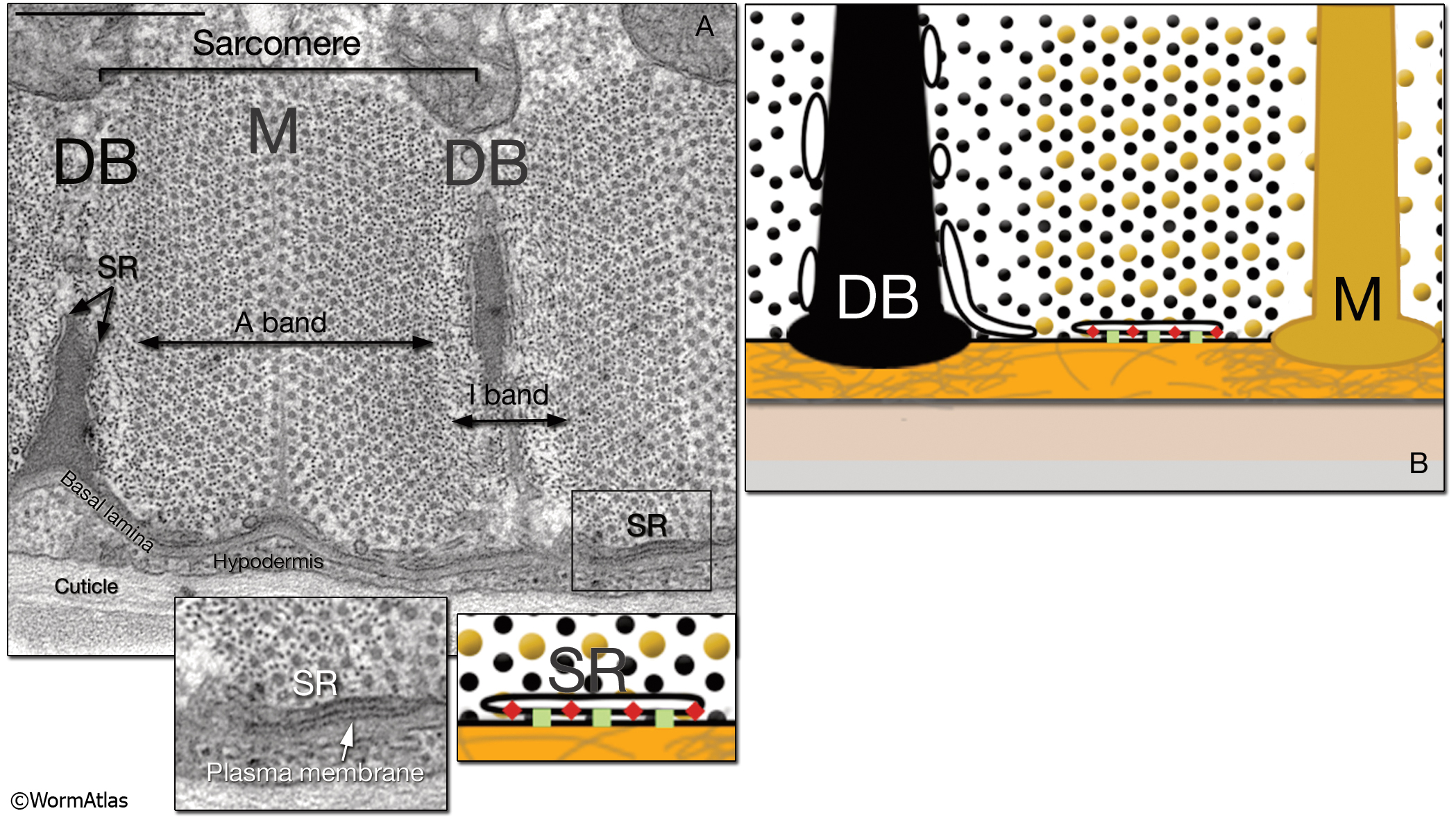
MusFIG 1A&B: The contractile apparatus in C. elegans.
A. Transmission electron microscopy (TEM) image of a cross section of the contractile apparatus in a body wall. The filaments of the lattice are oriented longitudinally and perpendicular to the surface. Dense bodies (DBs) anchor the thin (actin) filaments, whereas M line-homologs anchor the thick (myosin) filaments. A single unit of myofilament lattice between two DBs is called a sarcomere and contains one A band in the middle and two juxtaposing half I bands. In C. elegans, each adult body wall sarcomere is about 1 μm wide. Bar, 0.5 μm. (SR) Sarcoplasmic reticulum. (Image source: [Hall] N501C R4.)
B. Diagram illustrating the contractile lattice and the placement and structure of the SR. The vesicular membranous network of SR surrounds the myofilament lattice and is present along dense bodies and the apical plasma membrane underneath the lattice (see left inset, A). (Green squares)Voltage gated Ca++ channels (EGL-19); (red oblongs) ryanodine receptors; (yellow dots) thick filaments; (black dots) thin filaments; (orange layer) basal lamina with extracellular matrix (gray filaments); (beige layer) hypodermis; (gray layer) cuticle
See also MusFIG 1C&D.
Click on picture for full resolution image.
|