|
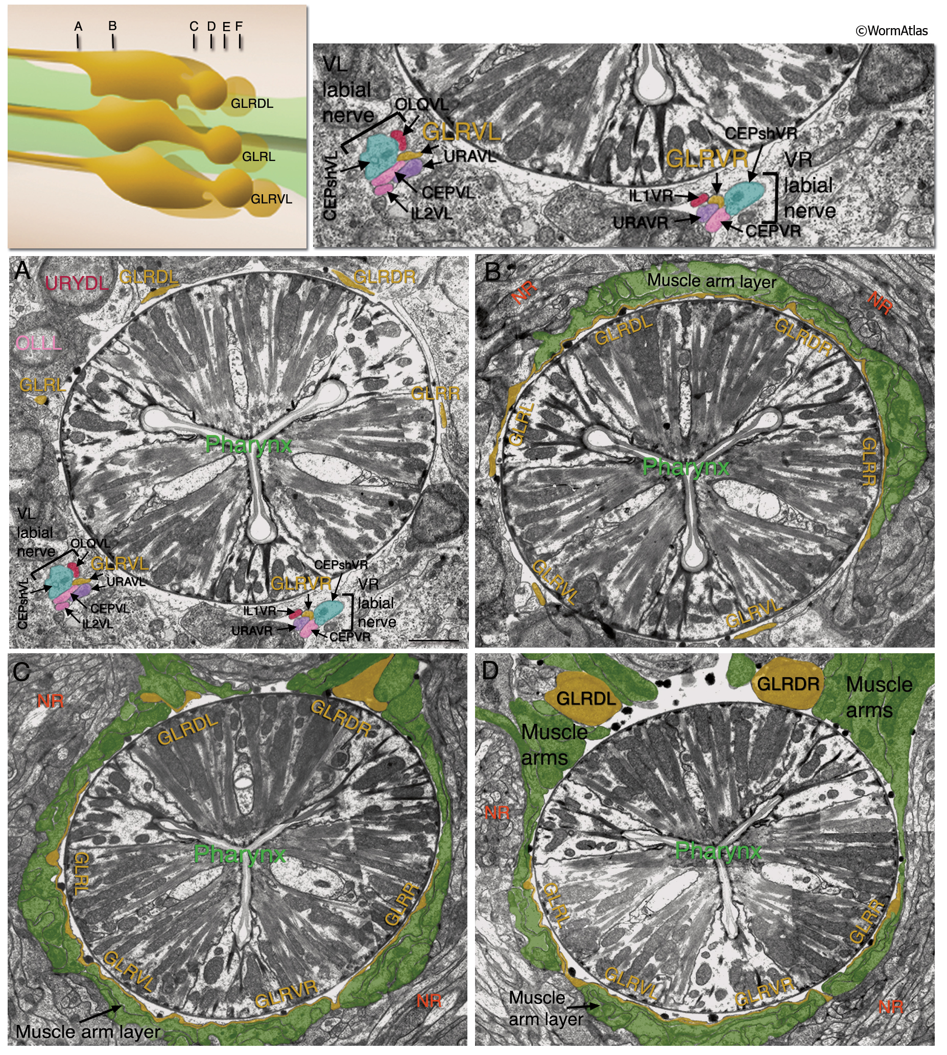
GlrFIG 3A-D: Fine structure of the GLR cells.
All except the top left inset are transverse-section TEMs. (NR) Nerve ring. (Top left inset) Section levels in A–F. Bars, 1 μm.
A. Anteriorly to the nerve ring, the thin GLR processes join the labial process bundles. At the level shown, only GLRVL/R processes have joined the labial bundles (same area is magnified in top right inset), whereas GLRL/R and GLRDL/R process bundles are still seen closer to the pharynx. Some neuron cell bodies of the anterior ganglion are labeled. The GLR processes contact RME processes to make gap junctions in this region (not shown). (Image source: N2U [MRC] A17517-20.)
B. Slightly posterior to the level in A, the muscle plate (muscle arm layer, green) is seen to be forming on the outside of the thin GLR processes, each of which occupies almost one-third the circumference of the isthmus. There is no BL between GLR cells and muscle arms. (Image source: N2U [MRC] A182 21-22.)
C. More posteriorly, the muscle plate wraps around the whole circumference of the isthmus, outside the thin GLR processes. Outside the muscle plate, the nerve ring processes are seen. A basal lamina (not shown) separates the muscle plate from the nerve ring. (Image source: N2U [MRC] A191 4-7.)
D. Posteriorly to the nerve ring, the first cell bodies that are seen are those of the dorsal pair of GLR cells. The muscle arms that turn inward to reach the muscle plate are in close contact with the cell bodies. (Image source: N2U [MRC] A195 9-12.)
See also GlrFIG 3E-I.
Click on picture for full resolution image.
|