|
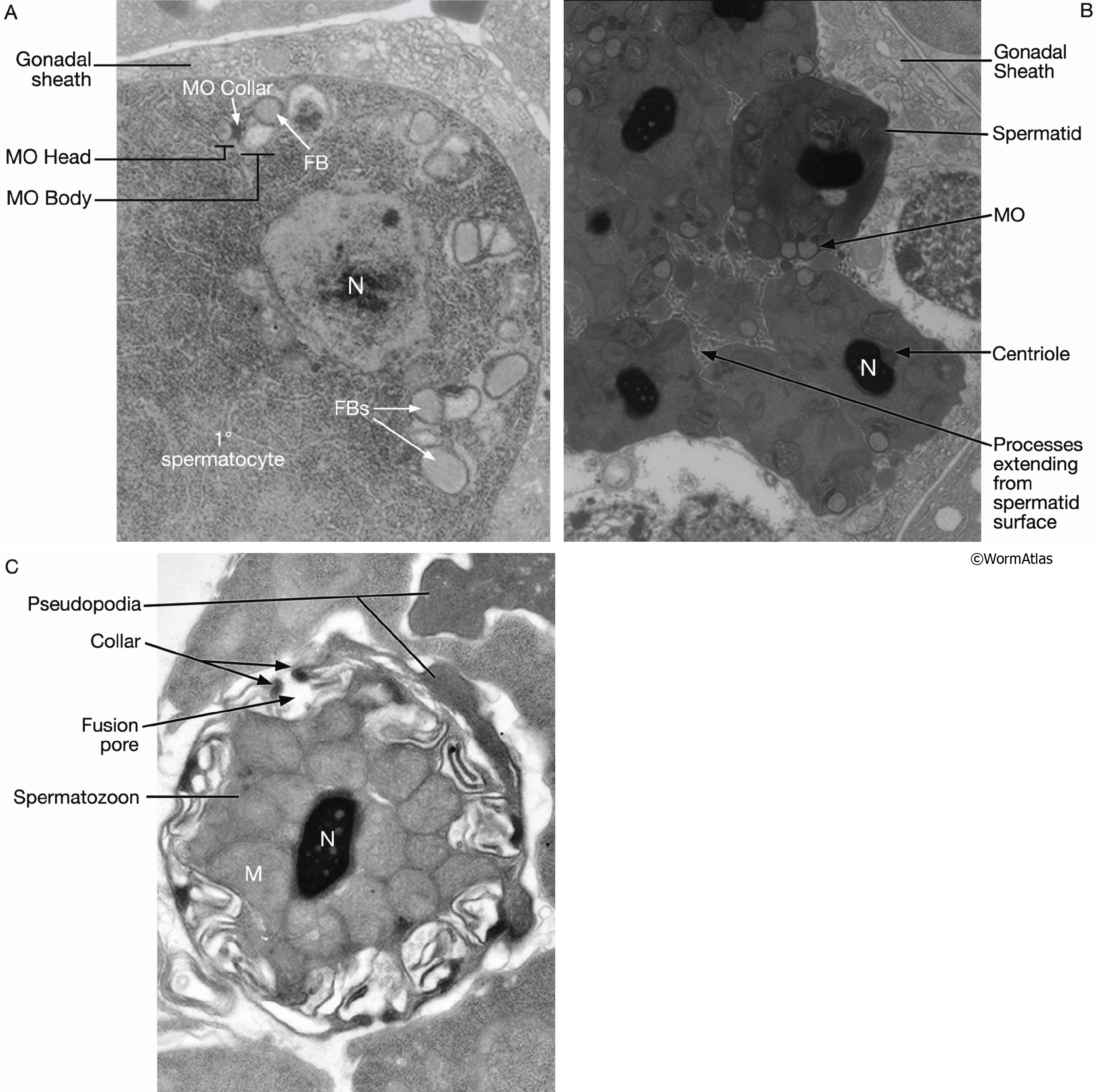
GermFIG 9: Electron micrographs of FB-MOs.
A. TEM, transverse section, of a late-L4 hermaphrodite (before first ovulation), showing FB-MOs beginning to assemble in a primary spermatocyte. (N) Nucleus. (Image source: N506 [Hall] Y810.)
B. TEM, transverse section, from a more proximal region of the animal in A, showing spermatids containing late-stage MOs, devoid of FB and collecting at the cell membrane. (N) Nucleus. (Image source: N506 [Hall] Y858.)
C. TEM, transverse section, of a spermatozoon showing a pore formed by fusion of an MO with the cell membrane. (M) Mitochondria. (Image source: [Hall] H707.)
Click on picture for full resolution image.
|