|
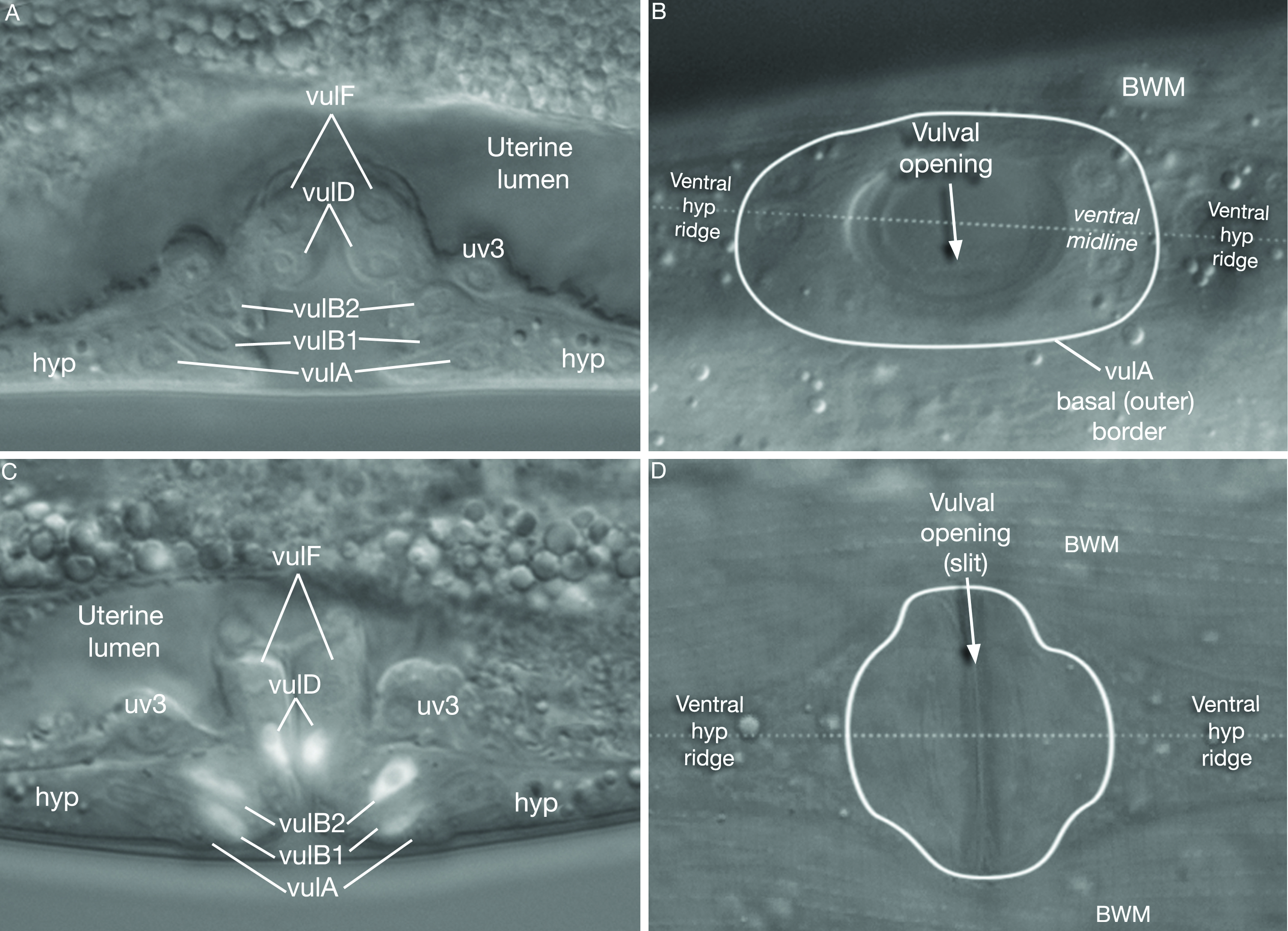
EggFIG 10: Vulval morphology before and after eversion.
A. DIC image of a mid- to late-L4 animal, before eversion, lateral view, midsublateral plane.
B. DIC image of a mid L4 animal, before eversion, ventral surface view, showing the vulval opening along the ventral midline.
C. DIC/epifluorescent image of a lin-11::GFP transgenic late-L4 animal, post-eversion, lateral view, midsublateral plane. (Strain source: B.P. Gupta and P.W. Sternberg.)
D. DIC image of a young adult animal, post-eversion, ventral surface view, showing the vulval slit.
Click on picture for full resolution image.
|