|
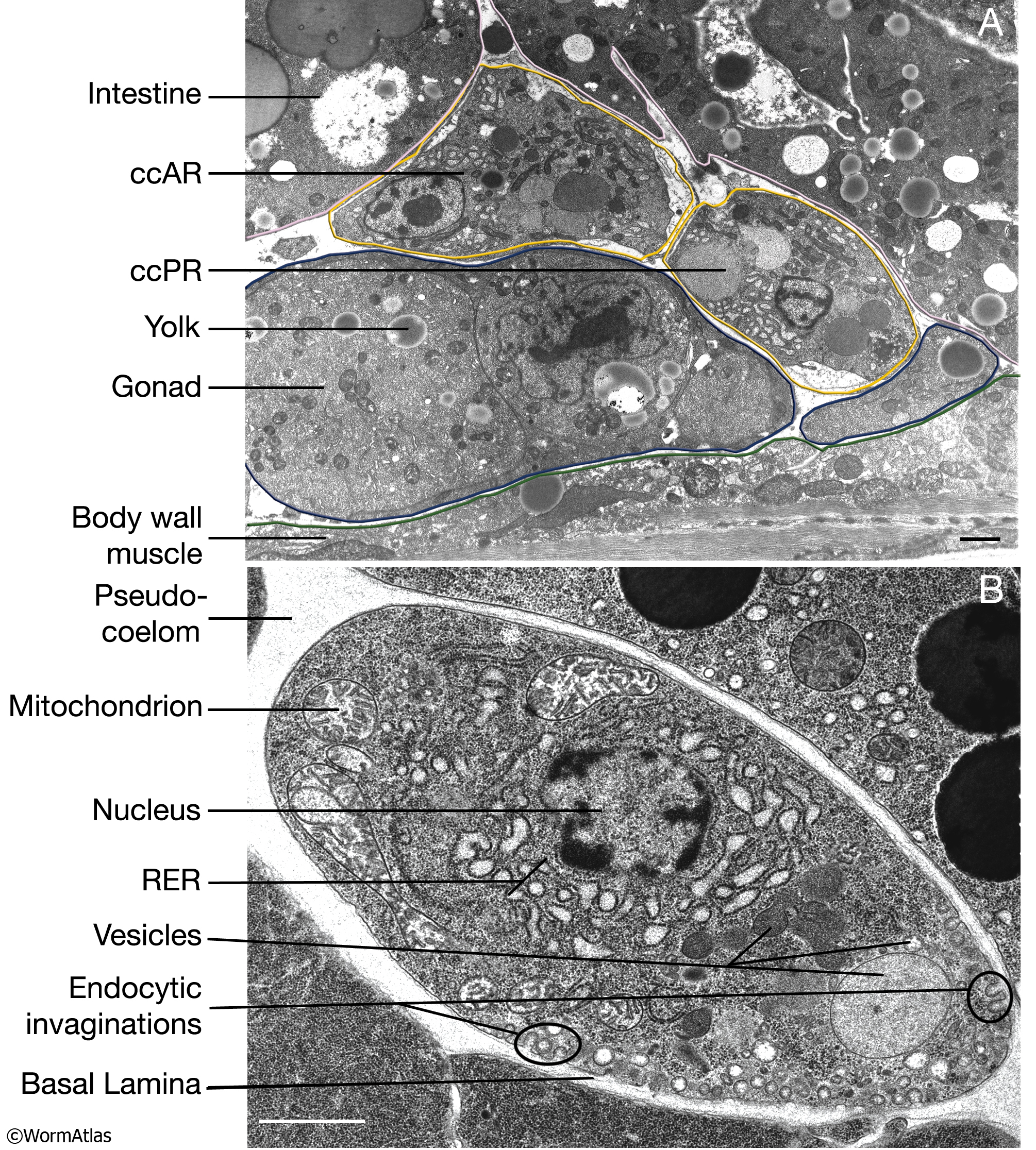
CcFIG 5: Electron micrographs of coelomocytes.
A. Transmission electron micrograph (TEM) of ventral right coelomocytes in an adult animal. The cells are located in the pseudocoelomic space between the body wall muscle, intestine and gonad. Foreign substances in the body cavity reach the cc’s by the movement of the pseudocoelomic fluid during animal’s locomotion and are endocytosed by these cells. Coelomocytes do not take up yolk particles from pseudocoelom that are made by the intestine; yolk particles are endocytosed by the neighboring gonad instead. Bar, 1 μm. (Image source: [Hall] N533-negative N543.
B. TEM of an individual coelomocyte. Multiple invaginations of plasma membrane are seen in regions where active endocytosis is taking place. Material is transported from these early endosomes to either the recycling endosomes, to be redirected to the cell surface, or late compartment endosomes and lysosomes, to be degraded. These compartments are seen as various sized vesicles within the cell (Fares and Grant, 2002; Treusch et al., 2004). The coelomocyte is surrounded by its basal lamina. Bar, 1 μm. (Image source: [Hall] negative F777.) |