|
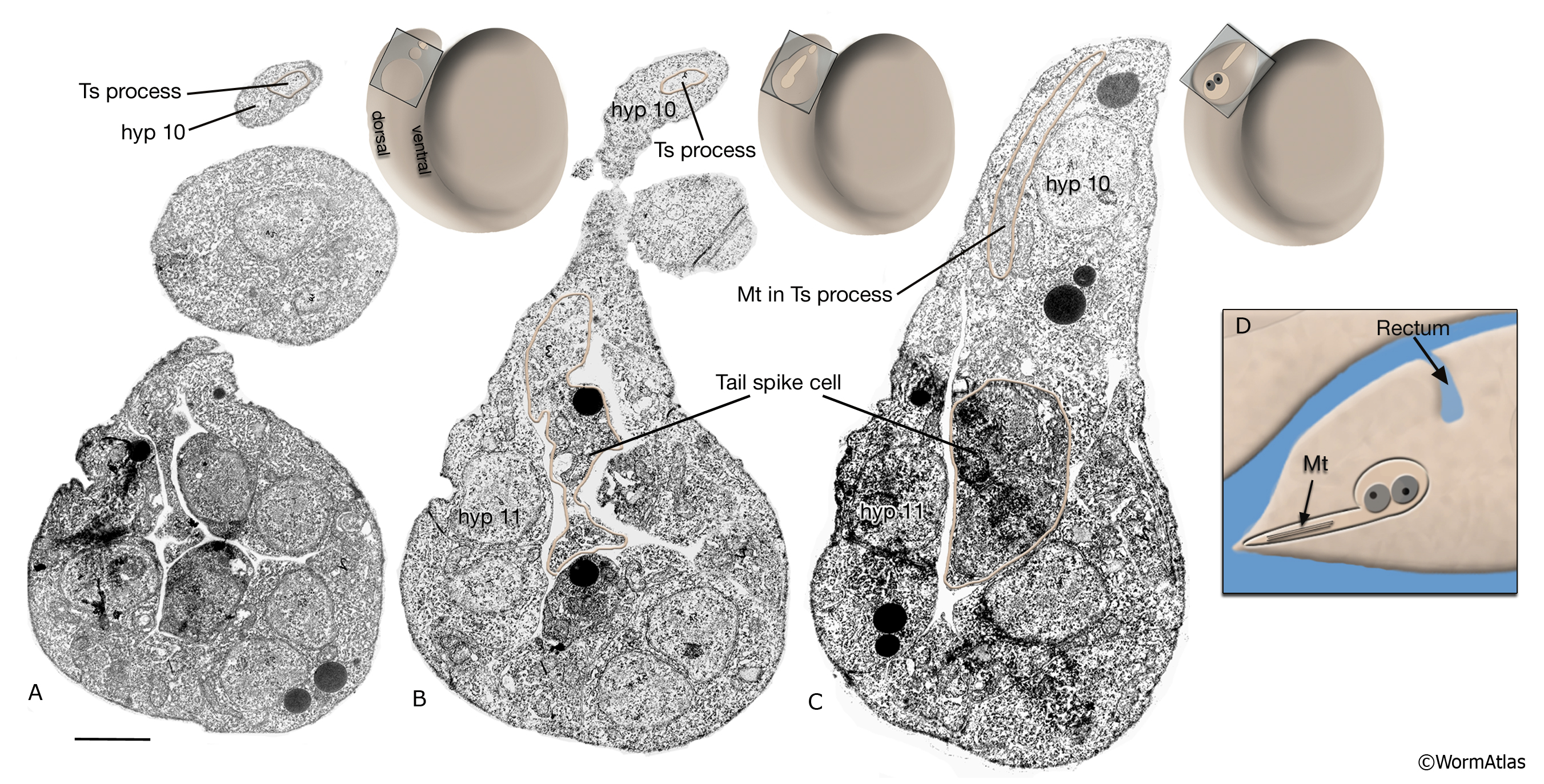
AtypFIG 4: Tail spike cells in twofold embryo.
A-C. TEMs, caudal at top. Longitudinal sections through the tail from lateral (A) to median (C) planes. (Insets) Levels of the sections. The tail spike plasma membrane is outlined in pink. Tail spike process (Ts) extends to the tip of the tail (A,B). The tail spike soma with two nuclei is located at midline (C). The microtubule (Mt)-containing process extends posteriorly from the soma. At a later stage in embryogenesis, the tail spike cell undergoes programmed cell death. Bar, 1 μm. (Image source: [MRC] Twofold embryo, [A] A997-6a, [B] A997.2, [C] A996-22.) .
D. Schematic representation of the syncytial tail spike cell and its process, lateral view. (Mt) microtubules.
Click on picture for full resolution image.
|