|
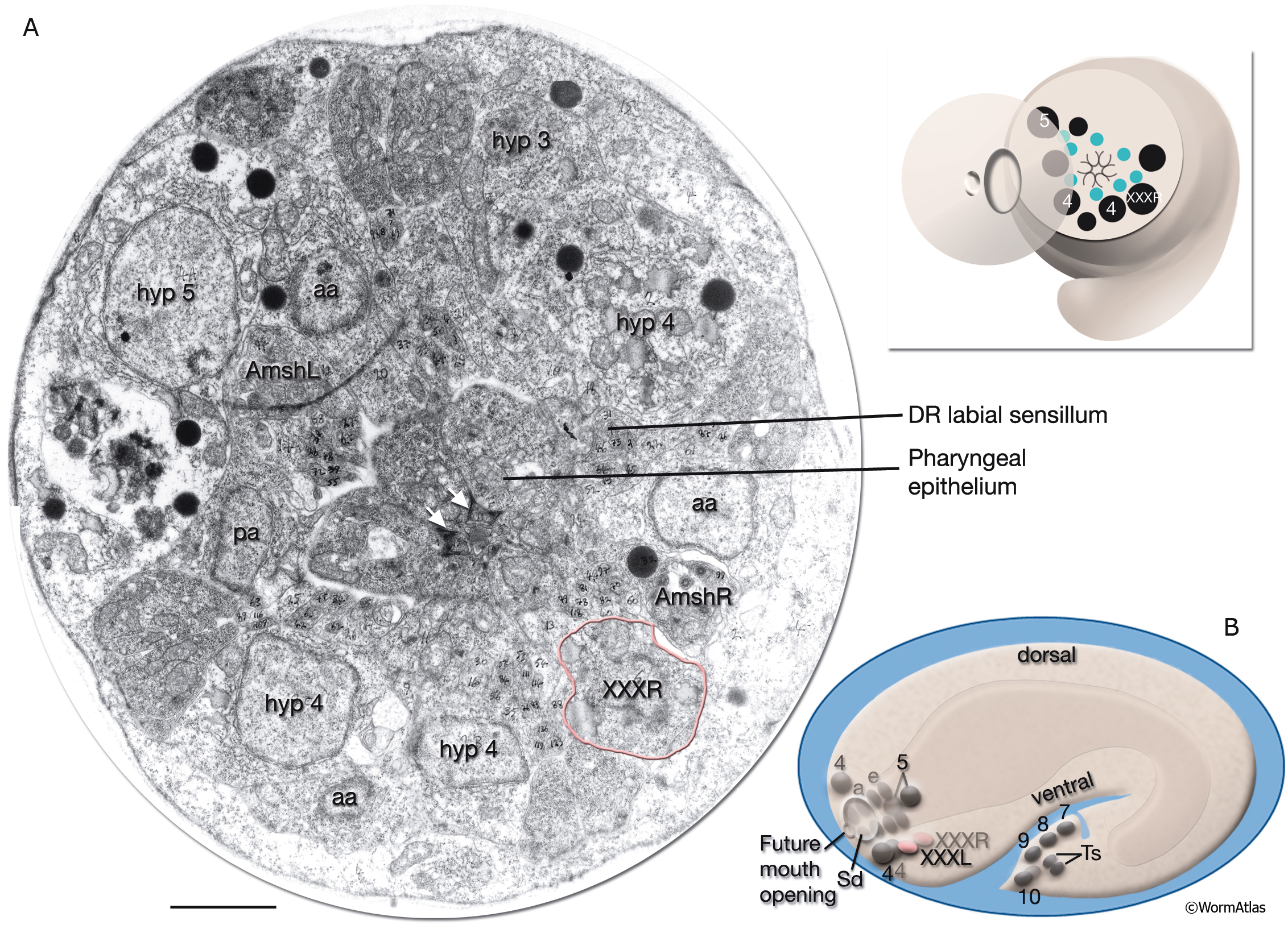
AtypFIG 1: Hypodermal origin of XXX in early embryo.
A. TEM, transverse section of the head at the level of XXXR soma, dorsal is top. (Inset, top) The level of the section. The cell bodies of hypodermal and arcade cells are aligned along an outside ring, whereas developing labial and amphid sensilla are located in an inner ring. Pharyngeal epithelium makes a tubular structure (without lumen at this stage) in the middle. (aa) Anterior arcade cells; (pa) posterior arcade cell; (white arrows) adherens junctions between pharyngeal epithelial cells; (green dots [inset, top]) labial and amphid sensilla; (black dots) hypodermal, arcade and XXXR nuclei. Bar, 1μm. (Image source: [MRC] Tadpole stage A1009-15.)
B. Schematic representation of the embryo as seen from the left side. At this stage, XXXL/R cells (pink ovals) are found near hyp 4 cells (4) on the ventral side of the embryo’s head after migrating from the dorsal side of the embryo where they are born and now lie in symmetric positions to hyp 5 cells (5). (Sd) Anterior sensory depression; (a) arcade cells; (e) pharyngeal epithelial cells; (other numbers) hypodermal nuclei in tail.; (Ts) tail spike cells.
Click on picture for full resolution image.
|