|
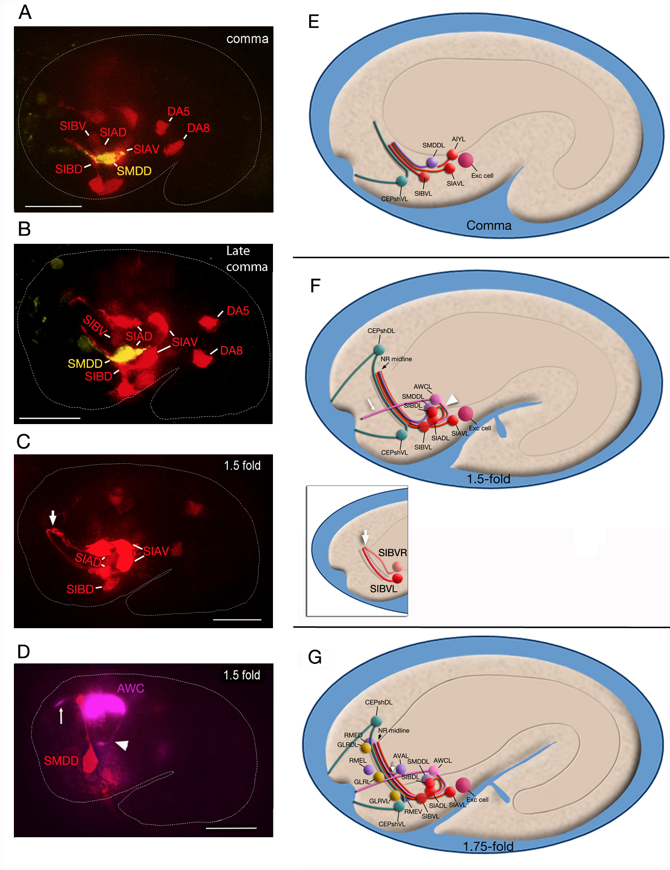
EmbryoNRDevFIG 3: The initiation and formation of the nerve ring
A-D. Fluorescent images of embryos corresponding to the stages shown in the right hand column. Scale bar: 10 µm.
E-G.Graphic images of nerve ring development in different stage embryos. Cell colors follow the WA color code.
A-C. Pceh-17::GFP marker; SIA, SIB, DA5, DA8 pseudocolored red, Pttx-3::mCherry marker; SMDD pseudocolored yellow.
D. Pttx-3::mCherry marker; SMDD, Phlh-16::GFP marker, AWC pseudocolored dark pink.
A&E. Comma stage embryo. Sublateral commissure neuron processes navigate anteriorly towards CEPshV and bundle with the dorsally-directed processes of these glia to position and initiate the nascent NR (among the sublaterals, SIA and SIB neuron processes grow slightly earlier than SMD processes and may be sufficient for this function)
C&D, F. 1.5-fold embryo. CEPsh and sublateral neuron axons cooperatively drive the follower processes (e.g. AIY, amphids) to enter the NR. White arrowhead, AWC amphid process which follows sublaterals into the nascent NR; white thin arrows, AWC dendrite growing towards the lip, white arrows, opposite side sublateral axons that reach each other at the NR midline by this stage
G. 1.75-fold embryo. In the older embryo muscles have migrated and separated into dorsal and ventral quadrants opening direct acess to the NR from the lateral ganglia. The time of muscle separation correlates with the extension of the first process (white arrow) in the lateral route (likely that of AVA neuron) (C. Norris and D.H. Hall, unpublished observations, 1997). While the mesoglial GLRs line the interior of the NR in adults, in the embryo, they are aligned around the posterior border of the NR while RME neurons surround the peripheral border of the NR (Rapti et al., 2017; C. Norris and D.H. Hall, unpublished observations).
Click on picture for full resolution image.
|