|
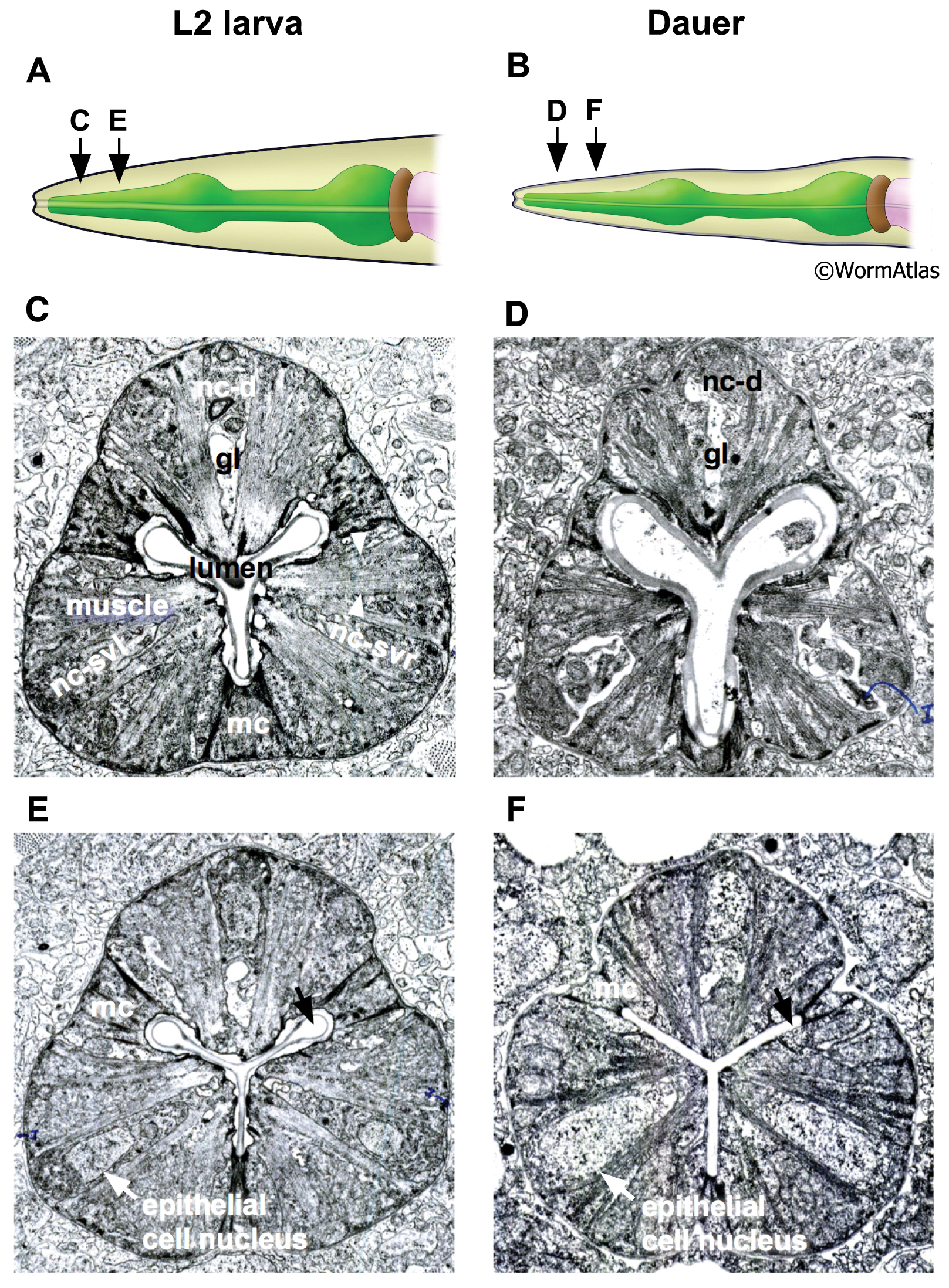
DPhaSUPFIG 2: Procorpus pharynx muscles.
A, C, E. Pharynx of L2 larva.
B, D, F. Pharynx of dauer larva. Two sections of the procorpus in an L2 and dauer are shown.
A&B. Arrows indicate approximate position of anterior (C&D) and medial (E&F) sections. Several structures are labeled in L2 sections, for comparison in dauer sections. Abbreviations: mc, marginal cell; nc-d, dorsal nerve cord; nc-svr, right subventral nerve cord; nc-svl, left subventral nerve cord. White arrowheads indicate m3 muscle fibers, which are shorter and more sparse in dauers. Black arrows indicate lumenal canals in the medial procorpus, which are closed in dauers. (Image sources: N2 L2 28-14 [D. Riddle] 175 and 236; N2 starved dauer 50-6-2 [D. Riddle] 293 and 40M.)
Click on picture for full resolution image.
|