|
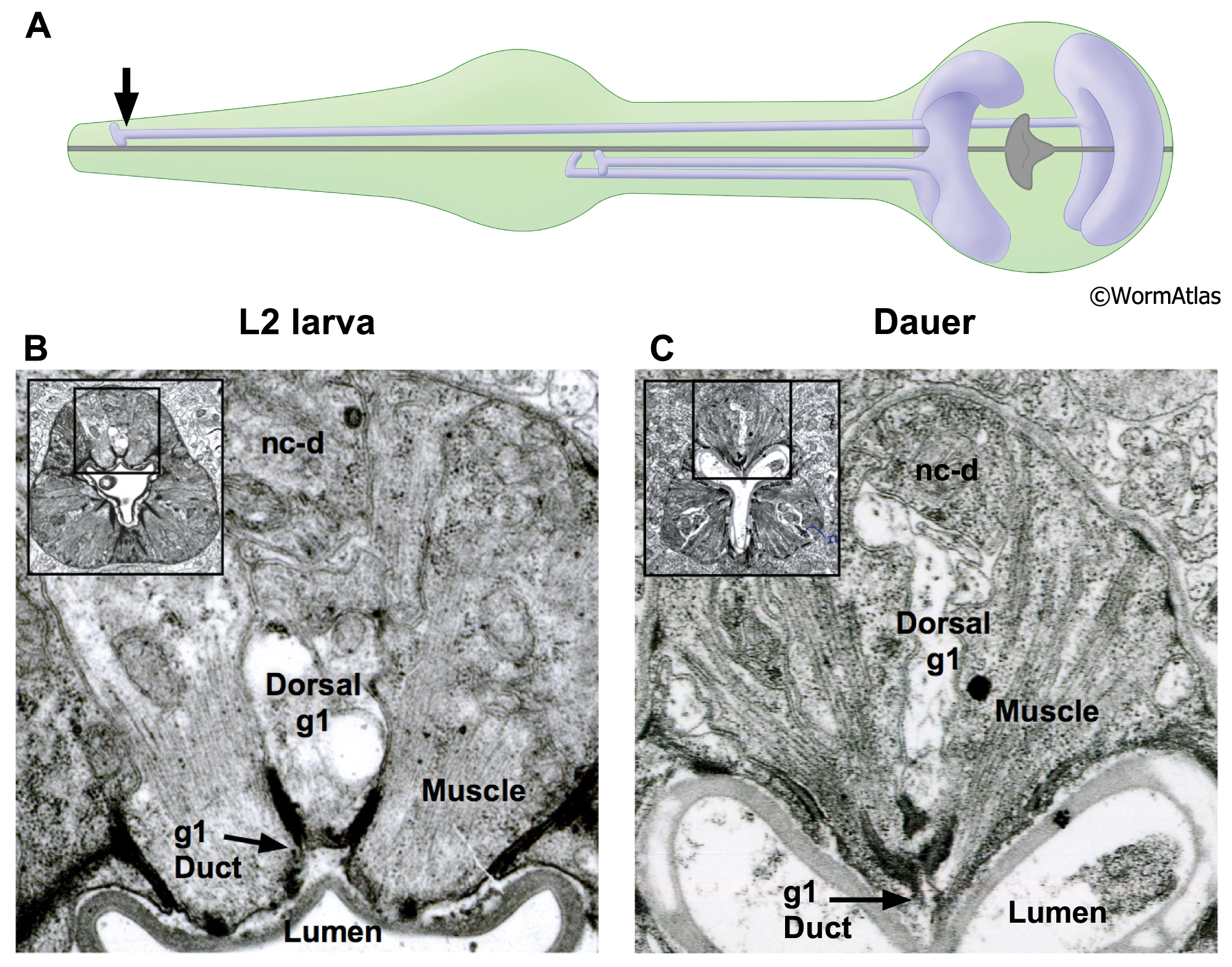
DPhaFIG 8: Dorsal g1 gland cell duct.
A. Illustration showing positions of dorsal and two ventral g1 gland cells in pharynx. Arrow indicates approximate position of sections in lower panels.
B. Transverse TEM section of anterior pharynx in L2 showing dorsal g1 gland (g1) in the vicinity of the cuticle-lined duct joining the gland process to the pharynx lumen. Insets show position of enlarged views on intact pharynx images. The dorsal nerve cord is indicated (nc-d). (Image source: N2 L2 28-14 [D. Riddle] 163.)
C. Transverse TEM section from dauer larva showing duct of dorsal g1 gland cell. (Image source: N2 starved dauer 50-6-2 [D. Riddle] 293.)
Click on picture for full resolution image.
|