|
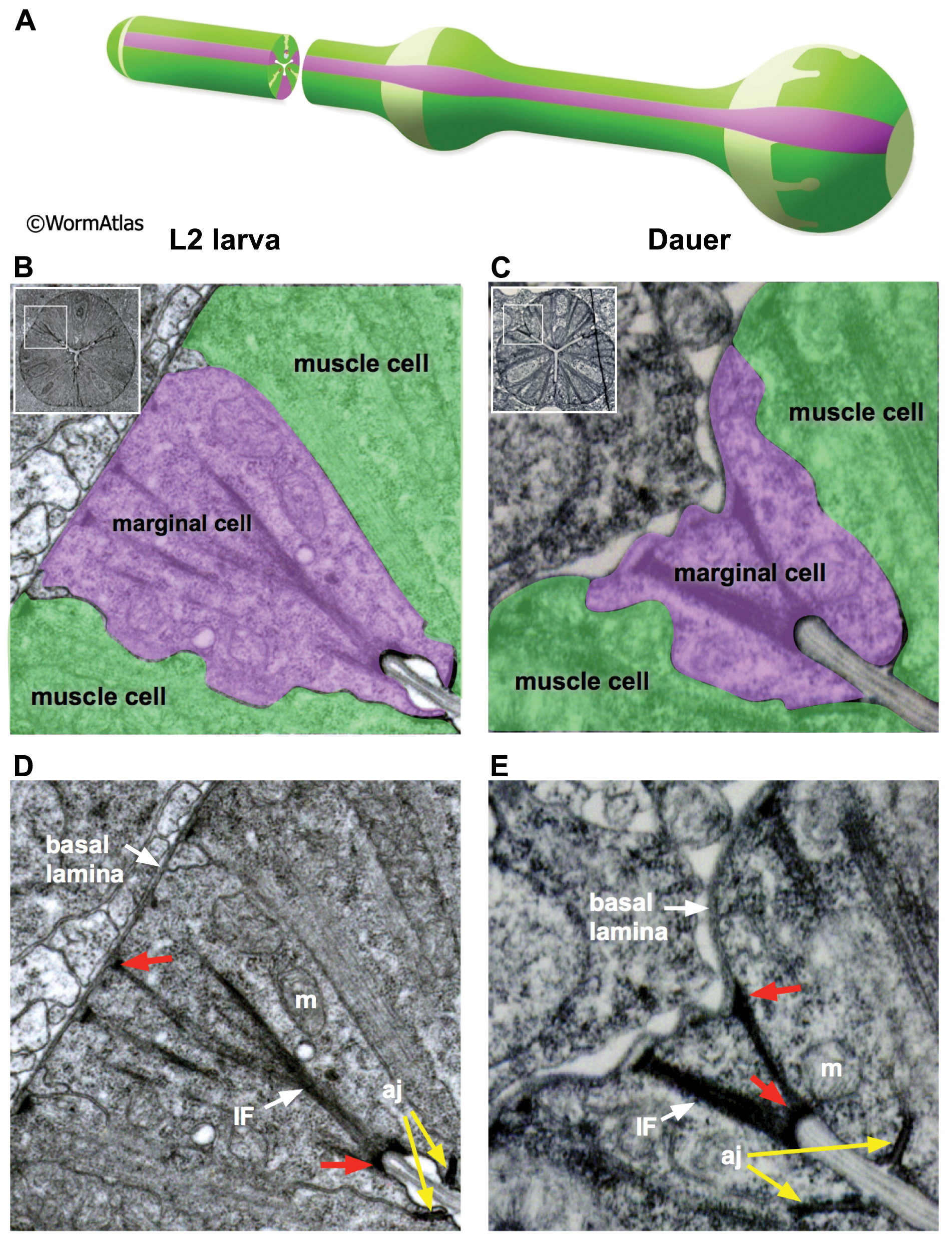
DPhaFIG 7: Marginal cells of the procorpus.
A. Illustration showing arrangement of marginal cells and muscle cells in the pharynx. (Image source: WormAtlas PhaFIG 7A.)
B&D. Transverse TEM section of marginal cells in an L2 larva.
B. Shading indicates different cell types. Muscle, green; marginal cells, purple. Inset indicates location of enlarged region within the intact pharynx.
D. Same image as in B, except without shading to show cellular details. (Image source: N2 L2 28-14 [D. Riddle] 436.)
C&E. Transverse TEM section of marginal cells in a dauer pharynx.
C. Shading as in panel B.
E. Same image as shown in panel C, without shading to show cellular details. (Image source: N2 starved dauer 50-6-2 [D. Riddle] 38M.)
D&E. Dauer marginal cells may contain thick intermediate filaments (IF) which are anchored by hemi-desmosomes (red arrows) between the apical membrane at the lumen and the basal lamina bordering the pseudocoelomic cavity. Mitochondria are visible in marginal cells of dauer and non-dauer larvae (m). At the apical membrane, the marginal cells are attached to neighboring muscle cells by adherens junctions (aj) linking them firmly to the pm muscles.
Click on picture for full resolution image.
|