|
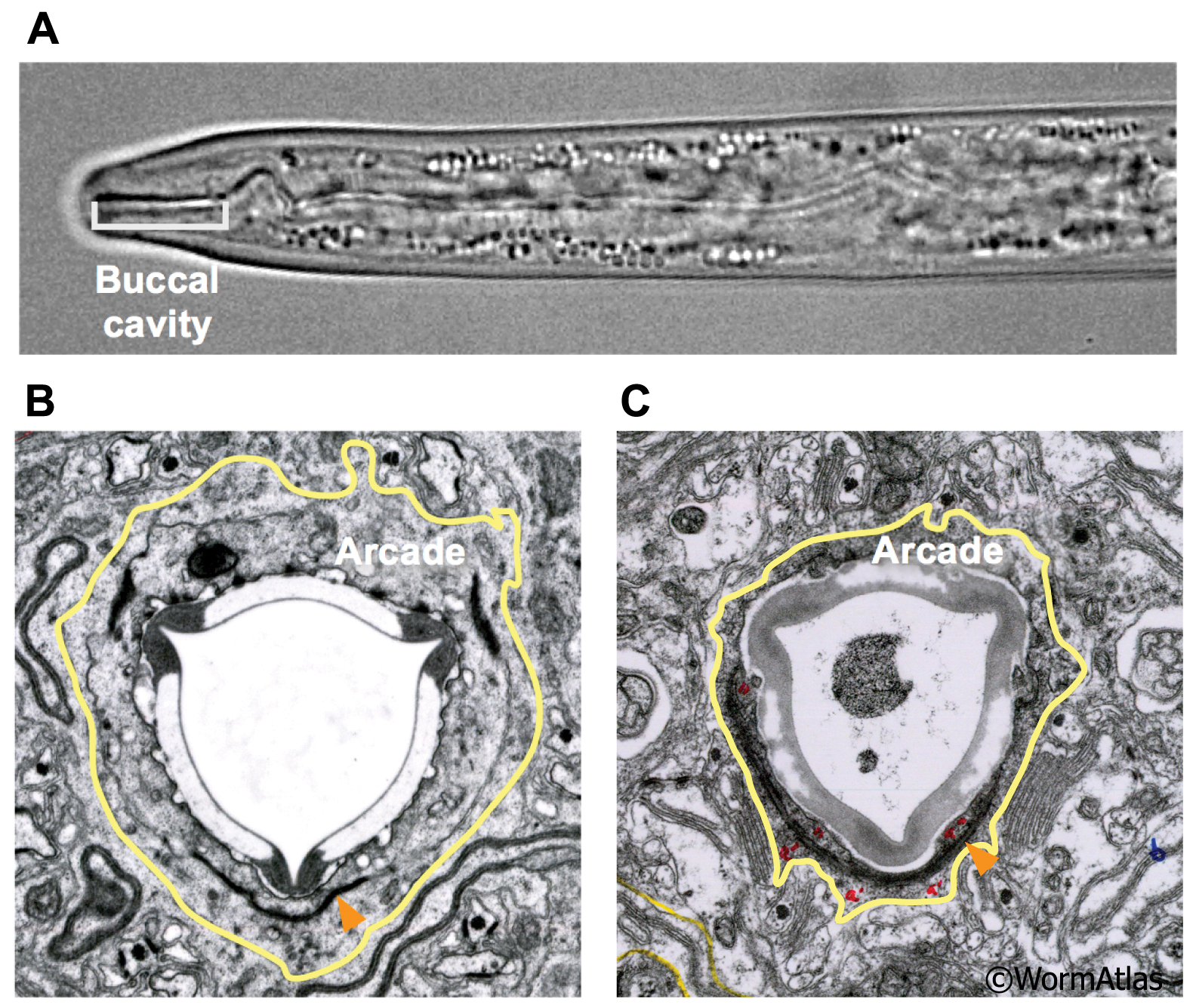
DPhaFIG 3: Arcade epithelium of dauer buccal cavity.
A. Dauer pharynx with buccal cavity indicated. (Image source: W.B. Iser, NIA.)
B. Transverse TEM section of non-dauer L4 showing arcade epithelium bordering the prostom cuticle. Yellow lines indicate position of arcade cell basal membrane. The apical edge borders the prostom cuticle. (Image source: N2_L4 recovered from dauer [D. Riddle] 11.)
C.Transverse TEM section showing arcade epithelium in dauer buccal cavity with basal edges outlined in yellow as for (B). Arrowheads indicate adherens junctions connecting anterior and posterior arcade. Red letters designate anterior arcade (a') and posterior arcade (a", "). (Image source: N2 starved dauer 50-2-1 [D. Riddle] 58.)
Click on picture for full resolution image.
|