|
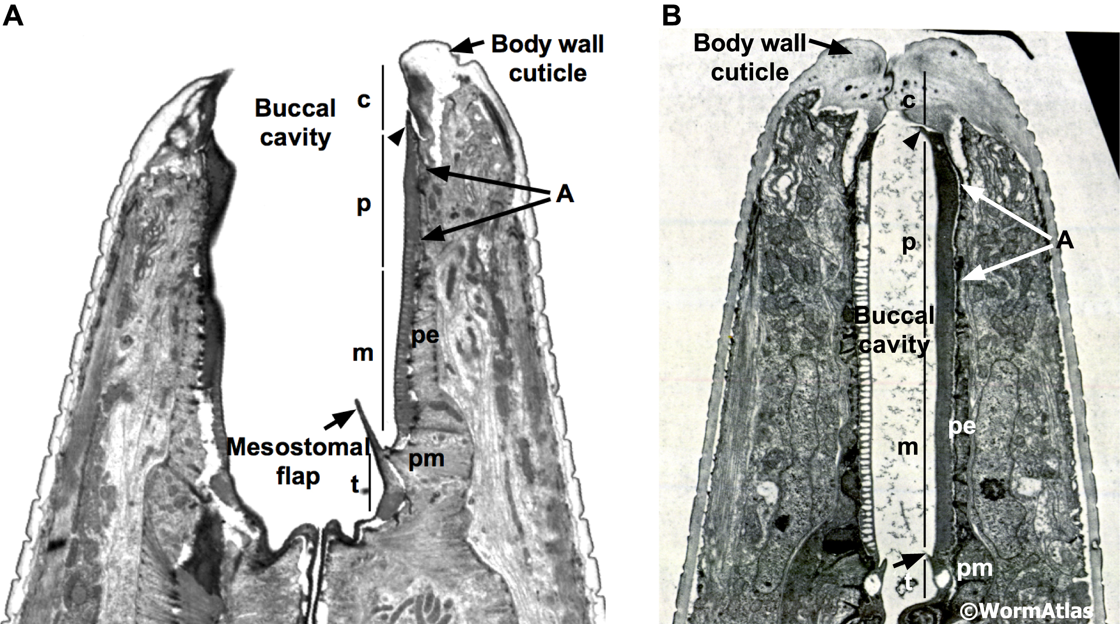
DPhaFIG 2: Buccal cavity in the dauer larva.
A. Horizontal TEM section through buccal cavity in an adult. The four regions of the buccal cavity are indicated: cheilostom (c), prostom (p), mesostom (m) and telostom (m). The cheilostom (c) cuticle is continuous with the body wall cuticle and has also been termed the bridging cuticle. The prostom cuticle (p) is produced by the anterior arcade and the posterior arcade cells (A). At the anterior, the prostom cuticle folds over the posterior region of the cheilostom cuticle (arrowhead). The mesostomal (m) cuticle is produced by the pharyngeal epithelium (pe) and the telostomal (t) cuticle is laid down by the anterior pharyngeal muscles (pm). The mesostomal flaps, which are located between the mesostomal and telostomal cuticles, are shown (short arrow). (Image source: B156 [D. Hall] lengthwise nose 7772.)
B. Horizontal TEM section through buccal cavity in a dauer larva. The dauer buccal cavity is occluded by a plug of body wall cuticle. The cheilostom (c) cuticle in the dauer is thicker and may extend slightly farther to the posterior. The extension of the cheilostom cuticle creates larger folds in the prostom cuticle (arrowhead). The mesostomal flaps appear to be smaller in the dauer than in the adult (short arrow). (Image source: N2 starved dauer 50-7-1 [D. Riddle] 43b.)
Click on picture for full resolution image.
|