|
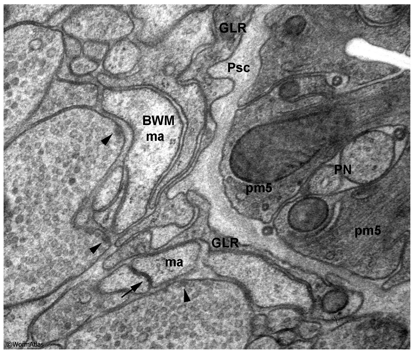
DNeuroFIG 9: Synapses in the dauer nerve ring.
Thin section from sample N974, prepared by high pressure freeze and freeze substitution showing both chemical and electrical synapses in the dauer. Arrowheads indicate neuromuscular junctions (chemical synapses) from two motor neuron axons onto the muscle arm of a bodywall muscle (BDW ma). Arrow indicates a gap junction (electrical synapse) between two other muscle arms (ma). Note that the typical chemical synapse shows a cluster of presynaptic vesicles close to a small electron dense bar on the presynaptic side, but rather little electron density, if any, on the postsynaptic membrane. A synaptic cleft separates the presynaptic axon from the postsynaptic muscle arm. The electrical synapse features two adjacent plasma membranes whose outer membrane leaflets are virtually touching and even in spacing. Psc; pseudocoelom. GLR; support cell for the muscle arms. PN; pharyngeal nerve. pm5; pharyngeal muscle cell.
(Image source: N2 N974 [K. Nguyen & D. Hall].)
Click on picture for full resolution image.
|