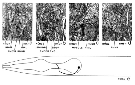
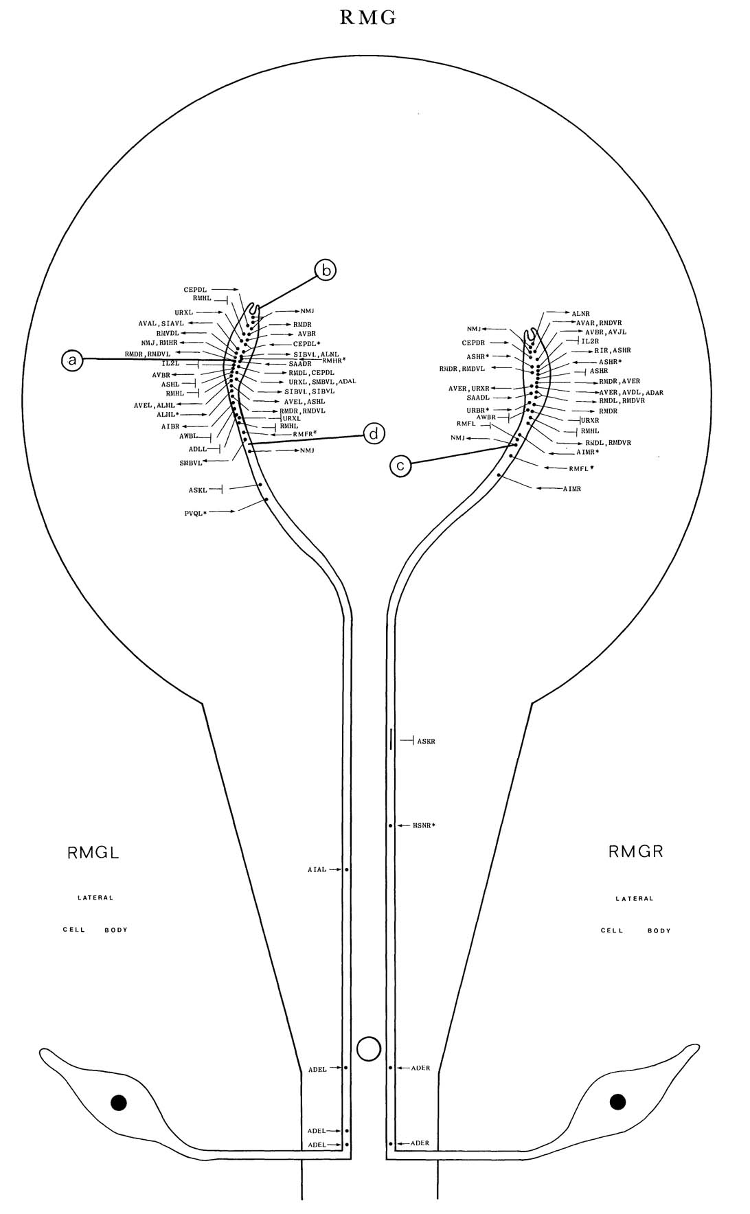
RMG is a set of two motoneurons, which innervate muscles in the head via NMJs in the
nerve ring. The cell bodies of RMG are situated laterally, just behind the pharynx, and enter
the ventral cord via the deirid commissures. The processes run near the dorsal surface of the
ventral cord and enter the nerve ring, which they run partly round, near the inner surface,
terminating sub-dorsally at characteristic structures, where they wrap round a bundle of
processes (b). RMG has NMJs on the lateral side of each of the four muscle spurs (c and figure
14). The synaptic vesicles are large; some are dark-cored (a). It also has synaptic output to RMD (a) and AVE. The main synaptic input is from ADE, CEP and also, possibly, RMF via processes that intercept NMJs (d). RMG has gap junctions with many classes of cell,
namely AWB (*d), RMH (*d), RMF (*h), ASK, IL2L/IL2R, ASH, ADL and URX.
Magnifications: (a) x 25500, (b-d) x 12750.


Click pictures for higher resolution images