Click pictures for higher resolution images Click pictures for higher resolution images
Members: DVA.
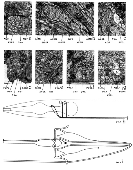
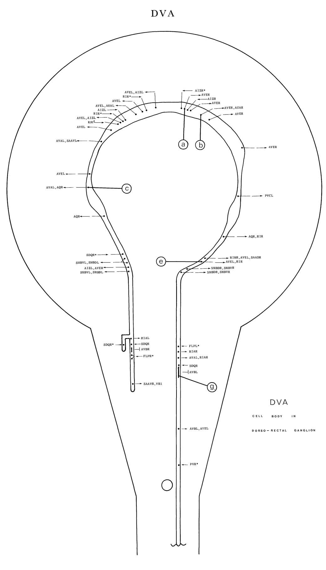
DVA is a single interneuron with its cell body situated in the dorso-rectal ganglion. An
anteriorly directed process enters the pre-anal ganglion (i) and runs in the ventral part of the
process bundle for the whole length of the ventral cord. It enters the nerve ring on the right-hand
side and travels right round it in an anticlockwise direction (h) running near the centre of the
neuropile in close association with the process of AQR; it then ends shortly after rejoining the
ventral cord on the left hand side. The process of DVA is generally rather large and has large,
vesicle-filled varicosities in the nerve ring (b). The vesicles tend to be irregularly shaped, except
in the vicinity of the presynaptic specializations, where they are smaller and more spherical
(a, b). In the nerve ring the main synaptic output is to AVE (a, b, e); there are some smaller
synapses to RIR (e), AVA (c), AQR (c), SMB, AIZ and AUA (b) and there are gap junctions
to AVB (g). There is a little synaptic input from AIZ and SDQ (*a). In the ventral cord there
are some small synapses to PVC (f), PVR, DBn (f) and VBn (d); there are several synapses
from PDE (*a) and some from PHC (*c), PLM (*e), PHA (*a) and PVD (*d); there are gap
junctions to PVR.
Magnifications: (a, b, d, g) x 25500, (c, e, f) x 17000.
DVA ventral cord synapses
partners |
gap junctions |
synapses from |
synapses to and corecipients |
PVC |
- |
3m |
2DB7, DB5, DB3 |
PDE |
- |
22+ 14 m |
1, VB11 |
PVR |
2 |
- |
1, DB2 |
DB3 |
- |
- |
1, PVC |
DB7 |
- |
- |
2PVC |
VB11 |
- |
- |
PDE, VA12 |
DB4 |
- |
- |
1 |
VA12 |
- |
- |
VB11 |
DB2 |
- |
- |
PVR |
DB5 |
- |
- |
PVC |
PHC |
- |
4+7m |
- |
PLM |
- |
5m |
- |
PHA |
- |
1+3m |
- |
PVM |
- |
2+1m |
- |
AVK |
- |
1 |
- |

|
|