Click pictures for higher resolution images Click pictures for higher resolution images
Members: AVG.
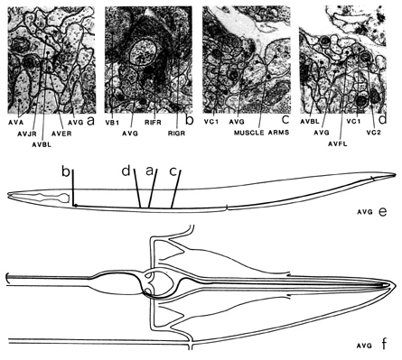
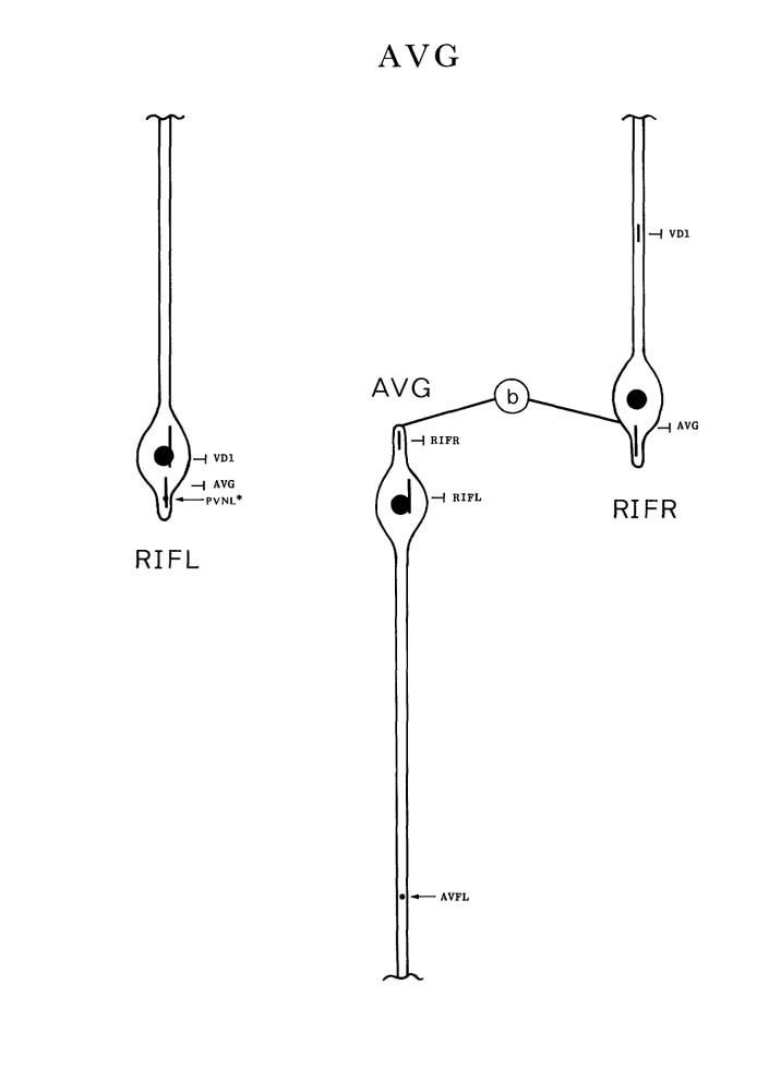
AVG is a single interneuron with its cell body situated in the retro-vesicular ganglion. A
posteriorly directed, fairly large process leaves the cell body and runs in the dorsal region of
the cord down to the pre-anal ganglion. Here it runs to the left of the anus and enters the
dorso-rectal ganglion and from there runs down the dorsal hypodermal ridge to the tip of the
tail. The disposition of the posterior extremities of this process suggest that it could be a sensory
dendrite. There are a few scattered synapses in the ventral cord (e.g. d) the most prominent
of which are some synapses to AVB (a). There are several synapses onto the basal lamina
surrounding the nerve cord with no obvious postsynaptic partners (c). The most striking
features of AVG are the gap junctions it makes with RIF in the retro-vesicular ganglion (b).
A short anteriorly directed process from AVG often pokes into the cell bodies of one of the RIF neurons (b).
Magnifications: (a) x 25500, (b-d) x 17000.
AVG ventral cord synapses
partners |
gap junctions |
synapses from |
synapses to and corecipients |
AVB |
- |
- |
3, AVJ |
AVA |
- |
- |
1, PVC, HSN |
PHA |
- |
2+6m |
PVQ, DA8 |
AVE |
- |
- |
AVE |
AVF |
- |
1+1m |
1 |
VA11 |
- |
- |
1 |
AVD |
- |
- |
1 |
DVB |
- |
- |
1 |
HDC |
- |
- |
1 |
DA8 |
- |
- |
PHA |
PVQ |
- |
- |
PHA |
PVC |
- |
- |
AVA |
AVJ |
- |
- |
AVB |
AVL |
- |
- |
PVP |
PVP |
- |
- |
AVL |
HSN |
- |
- |
AVA |
PQR |
- |
1 |
- |
RIF |
2 |
- |
- |
PVN |
- |
1m |
1, AVA |

|
|