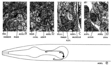
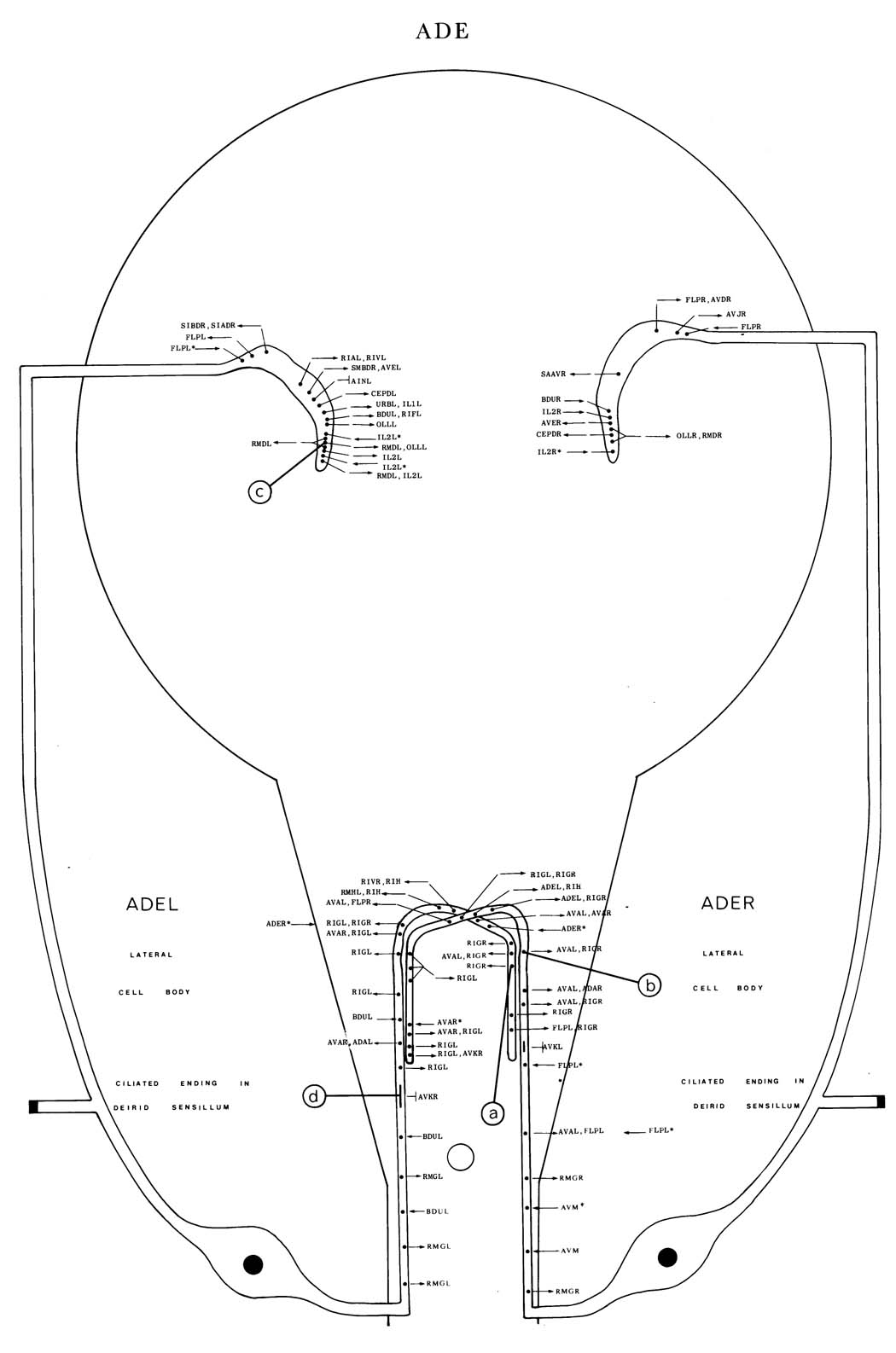
ADE is a set of two ciliated neurons with endings in the deirid sensilla, which are situated in the alae on the lateral lines. The cell bodies of ADE are part of a small group of cells situated laterally behind the second bulb of the pharynx. Processes enter the retro-vesicular ganglion via the deirid commissures and then run anteriorly in the ventral ganglion (e). Here they cross over to the contralateral side and run posteriorly for a short distance before ending. Most of the synaptic output is situated in this region and is predominantly to RIG (a) and RIG in association with AVA (b) although there is usually a bias towards RIG in these dyadic synapses. The process to the ciliated ending has a branch, which enters the ring neuropile laterally, running anteriorly through the ring neuropile and making some rather small synapses to diverse partners; OLL, RMD (c), CEP and FLP are the most prominent synaptic partners in this region. ADE receives some synaptic input from BDU (*b), FLP (*c), AVM (*) and IL2L/IL2R. It has gap junctions to AVK (d) in the neuropile of the ventral ganglion. ADE neurons have been shown to contain dopamine (Sulston et al. 1975). Magnifications: (a, c, d) x 25500, (b) x 12750.


Click pictures for higher resolution images