|
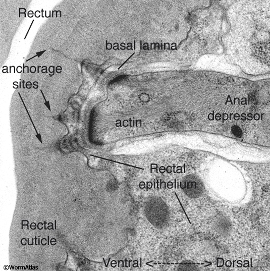
ETFIG 2: Electron tomogram of a rectal muscle.
A dual axis electron tomogram shows diffuse elements of the basal lamina and within the cuticle, including layers of filaments spanning the lamina, and long thin filaments anchoring the depressor muscle cell to the rectal cuticle. Ventral is to the left. This is not an electron micrograph. The tomogram has been resliced at a favorable angle. Such fine features are never seen well in standard thin sections. HPF/FS sample. Image capture by Leslie Gunther, FEI Technai20 TEM. Tomogram calculated using gold particles as reference marks.
This tomogram can also be viewed as a set of movies.
Click on picture for full resolution image.
|